Anesthesia: Types, Drug Classes, Monitoring & Safety Guide
Medical Governance: Authored by the Clinical Anesthesiology & Perioperative Medicine Editorial Board | Medically Reviewed by a Board-Certified Anesthesiologist & Critical Care Specialist | Verified Against ASA, ESAIC, WFSA, and AAGBI Clinical Practice Guidelines
Anesthesia is a specialized branch of perioperative medicine dedicated to the reversible pharmacological suppression of the nervous system. Far from merely putting a patient to sleep, modern anesthesia delivers four synchronized physiological endpoints, historically termed the anesthetic triad: reversible unconsciousness (hypnosis/amnesia), intense analgesia (suppression of pain perception), autonomic reflex stability (blunting sympathetic stress responses to surgical trauma), and skeletal muscle relaxation (paralysis).
Every year, hundreds of millions of surgical, diagnostic, and interventional procedures are performed safely worldwide under anesthesia. The specialty encompasses four major clinical modalities: general anesthesia, regional anesthesia (neuraxial and peripheral nerve blocks), monitored anesthesia care / procedural sedation, and local infiltration.
Patient safety in contemporary anesthesiology has improved dramatically over the past four decades, transforming an environment once carrying significant procedural mortality into one of the safest domains in modern medicine. This transformation has been driven by standardized multi-parameter physiological monitoring (pulse oximetry, continuous capnography, end-tidal anesthetic gas analysis, processed electroencephalography), sophisticated airway devices (video laryngoscopy, supraglottic airways), and advanced pharmacological agents engineered with rapid clearance profiles and specific, rapid-acting chemical reversal agents.
This clinical guide provides a comprehensive overview of anesthetic neurobiology, classifications, core pharmacological drug classes, preoperative evaluation and fasting guidelines, intraoperative monitoring standards, emergency crisis protocols, and post-anesthesia care.
Anesthesia Medicine Clinical Matrix
-
Intravenous Hypnotic Induction Agents:
-
Key Molecules: Propofol, Etomidate, Ketamine, Methohexital.
-
Mechanism of Action: Propofol and etomidate bind selectively to beta subunits of gamma-aminobutyric acid type A (GABA-AGABAA) receptors in the central nervous system, enhancing chloride ion influx to hyperpolarize postsynaptic neuronal membranes. Ketamine non-competitively antagonizes N-methyl-D-aspartate (NMDA) receptors, interrupting thalamocortical sensory integration.
-
Clinical Indications: Rapid induction of general anesthesia, maintenance of total intravenous anesthesia (TIVA), procedural sedation.
-
Critical Precautions & Monitoring: Vasodilation and direct myocardial depression causing systemic hypotension (propofol); transient adrenocortical suppression via 11-beta-hydroxylase inhibition (etomidate); emergence delirium, sympathetic tachycardia, and hypertension (ketamine).
-
Inhalational Anesthetic Volatile Agents:
-
Key Molecules: Sevoflurane, Desflurane, Isoflurane, Nitrous Oxide.
-
Mechanism of Action: Augments inhibitory GABAA and two-pore domain potassium channels (K2P) while suppressing excitatory synaptic receptors (nicotinic acetylcholine, AMPA, NMDA) across the brain and spinal cord, inhibiting conscious perception and immobilizing motor reflexes.
-
Clinical Indications: Maintenance of general anesthesia in operating suites; smooth, non-pungent mask inhalation induction in pediatric populations (sevoflurane).
-
Critical Precautions & Monitoring: Dose-dependent systemic vasodilation, drop in mean arterial pressure, coronary steal (isoflurane), airway irritation/coughing (desflurane); absolute trigger for Malignant Hyperthermia; monitor End-Tidal Minimum Alveolar Concentration (MAC).
-
Depolarizing Neuromuscular Blocking Agents (NMBAs):
-
Key Molecules: Succinylcholine (Suxamethonium).
-
Mechanism of Action: Binds directly to nicotinic acetylcholine receptors (nAChRs) at the neuromuscular junction motor endplate, triggering sustained muscle depolarization (visible fasciculations) followed by a refractory state that prevents further muscle action potentials.
-
Clinical Indications: Rapid Sequence Induction and Intubation (RSII) for emergency full-stomach airways, immediate laryngospasm relief.
-
Critical Precautions & Monitoring: Risk of acute hyperkalemic cardiac arrest in denervating neurological conditions, major burns, or massive crush injuries; prolonged paralysis in patients with plasma cholinesterase (pseudocholinesterase) deficiency; potent trigger for Malignant Hyperthermia.
-
Non-Depolarizing Neuromuscular Blocking Agents:
-
Key Molecules: Rocuronium, Vecuronium, Cisatracurium, Atracurium.
-
Mechanism of Action: Competitively antagonizes nicotinic acetylcholine receptors at the post-junctional motor endplate without triggering initial depolarization, blocking endogenous acetylcholine binding and causing flaccid skeletal muscle paralysis.
-
Clinical Indications: Endotracheal intubation, continuous surgical muscle relaxation for abdominal and thoracic operations, mechanical ventilator synchrony in intensive care.
-
Critical Precautions & Monitoring: Quantitative Train-of-Four (TOF) peripheral nerve monitoring; prolonged action in hepatic/renal clearance failure (rocuronium/vecuronium); cisatracurium undergoes organ-independent Hofmann elimination, making it preferred in renal/hepatic failure.
-
Neuromuscular Reversal Agents:
-
Key Molecules: Sugammadex, Neostigmine (paired with Glycopyrrolate).
-
Mechanism of Action: Sugammadex is a modified gamma-cyclodextrin that physically encapsulates steroidal NMBAs (rocuronium > vecuronium) in a 1:1 molecular complex in blood plasma, rapidly restoring neuromuscular function; neostigmine inhibits acetylcholinesterase, flooding the motor endplate with acetylcholine to outcompete non-depolarizing relaxants.
-
Clinical Indications: Rapid reversal of neuromuscular blockade at the conclusion of surgery, emergency reversal of deep rocuronium-induced paralysis.
-
Critical Precautions & Monitoring: Sugammadex binds hormonal contraceptives (requires backup barrier contraception for 7 days post-administration); neostigmine requires an anticholinergic (glycopyrrolate) to prevent profound muscarinic bradycardia and bronchospasm.
-
Amide and Ester Local Anesthetics:
-
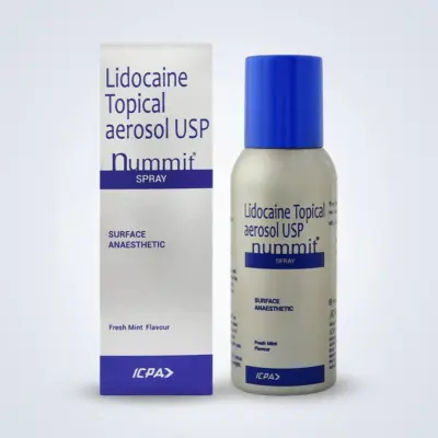
Key Molecules: Bupivacaine, Ropivacaine, Lidocaine, Mepivacaine, Tetracaine.
-
Mechanism of Action: Binds intracellularly to the S6 segment of domain IV on voltage-gated sodium channels (NaV), physically occluding the pore and preventing sodium influx. This halts action potential generation and propagation along sensory, autonomic, and motor nerve fibers.
-
Clinical Indications: Infiltration anesthesia, minor surgical excisions, regional peripheral nerve blocks, spinal (subarachnoid) and epidural neuraxial anesthesia, systemic anti-arrhythmic and multimodal analgesia infusions (IV lidocaine).
-
Critical Precautions & Monitoring: Local Anesthetic Systemic Toxicity (LAST); bupivacaine exhibits high cardiotoxicity (ventricular arrhythmias, refractory arrest); strict maximum weight-based dosing; aspirate prior to injection to avoid accidental intravascular delivery.
Neurobiology and Mechanisms of Anesthesia
Anesthetics do not target a single “consciousness switch” in the brain. Rather, they interrupt communication across distributed neural networks, preventing the brain from integrating sensory information:
1. Central Mechanisms of Hypnosis and Amnesia
General anesthetics act on several major anatomical centers within the brain:
-
The Cerebral Cortex and Thalamus: Anesthetics disrupt thalamocortical loops that relay information between sensory organs and the cerebral cortex. This causes functional fragmentation—the cortex continues to show isolated local electrical activity, but it can no longer share or integrate information across different brain lobes, resulting in unconsciousness.
-
The Ascending Reticular Activating System (ARAS): Located in the brainstem, the ARAS releases excitatory neurotransmitters (acetylcholine, norepinephrine, histamine, orexin) to maintain wakefulness. Anesthetic agents depress ARAS firing while activating the ventrolateral preoptic nucleus (VLPO) of the hypothalamus, engaging natural slow-wave sleep pathways.
-
The Hippocampus: Anesthetics suppress long-term potentiation in CA1 pyramidal neurons of the hippocampus at concentrations far lower than those required to produce unconsciousness, ensuring anterograde amnesia and preventing memory formation during surgery.
2. Molecular Targets: GABA, NMDA, and Two-Pore Potassium Channels
At the microscopic level, anesthetic agents alter transmembrane ion channels:
-
GABA-A Receptors: The primary inhibitory neurotransmitter receptor in the central nervous system. Binding by propofol, etomidate, or volatile agents prolongs the opening of ligand-gated chloride channels. Inward chloride currents hyperpolarize the post-synaptic neuron, driving its membrane potential further below threshold and suppressing action potentials.
-
NMDA Receptors: Major excitatory ionotropic glutamate receptors. Dissociative agents like ketamine block the open pore of the NMDA channel, cutting off excitatory signaling.
-
Two-Pore Domain Potassium Channels (K2P): Volatile gases open leak potassium channels (TREK and TASK channels), allowing intracellular potassium to exit the cell. This hyperpolarizes neurons, suppressing motor reflexes in the spinal cord.
Major Types and Modalities of Anesthesia
Anesthetic techniques are selected based on surgical location, patient comorbidities, procedural duration, and recovery goals.
Spectrum of Anesthetic Techniques
| Anesthetic Modality |
Level of Consciousness |
Airway Management |
Typical Clinical Indications |
Primary Drug Classes Employed |
| General Anesthesia |
Complete drug-induced unconsciousness; unarousable to painful stimuli |
Controlled; Endotracheal Tube (ETT) or Supraglottic Airway (LMA) with mechanical ventilation |
Major abdominal, thoracic, cardiac, intracranial, prolonged or laparoscopic surgery |
IV induction hypnotics, volatile inhalational agents, opioids, neuromuscular blockers |
| Neuraxial Regional Anesthesia |
Fully conscious or co-administered light/moderate sedation |
Spontaneous; natural airway reflexes preserved |
Lower abdominal, pelvic, urological, orthopedic hip/knee, and obstetric procedures |
Amide local anesthetics (bupivacaine, ropivacaine) ± lipophilic opioids (fentanyl, morphine) |
| Peripheral Nerve Blocks (PNBs) |
Fully conscious or co-administered procedural sedation |
Spontaneous; natural airway reflexes preserved |
Upper/lower extremity surgeries, localized orthopedic repairs, postoperative regional analgesia |
Long-acting local anesthetics (ropivacaine, bupivacaine) ± adjuvants (dexamethasone, clonidine) |
| Monitored Anesthesia Care (MAC) |
Ranges from minimal anxiolysis to deep procedural sedation |
Spontaneous; patient typically maintains patent airway independently |
Endoscopies, colonoscopies, minor dermatological excisions, cataract extractions, catheterizations |
Short-acting IV sedatives (propofol, midazolam, dexmedetomidine) and analgesics (fentanyl) |
| Local Infiltration Anesthesia |
Fully conscious and alert; completely uncompromised |
Spontaneous; completely unaltered normal respiratory status |
Minor skin repairs, superficial wound suturing, punch biopsies, localized dental procedures |
Fast-acting local anesthetics (lidocaine, mepivacaine) ± vasoconstrictor epinephrine |
1. General Anesthesia
Indicated for major abdominal, thoracic, cardiac, intracranial, and laparoscopic surgeries:
2. Regional Anesthesia: Neuraxial Modalities
Involves injecting local anesthetics close to the spinal cord or spinal nerve roots, rendering large anatomical regions numb without causing unconsciousness:
-
Spinal (Subarachnoid) Anesthesia: Dense sensory, motor, and autonomic blockade from the mid-abdomen down to the toes. Ideal for Cesarean deliveries, total hip/knee replacements, and transurethral urological procedures.
Epidural Anesthesia:
-
Technique: A larger Tuohy needle is advanced into the epidural space (identified using the “loss-of-resistance” technique) without puncturing the dura. A thin plastic catheter is threaded into the space, allowing continuous or repeated infusions of local anesthetics and opioids.
-
Clinical Role: Slower onset (15 to 20 minutes) than spinal anesthesia, but permits titratable, extended analgesia over days, serving as the gold standard for labor analgesia and post-thoracotomy/major abdominal pain control.
3. Regional Anesthesia: Peripheral Nerve Blocks (PNBs)
Under real-time high-resolution ultrasound visualization, local anesthetic is deposited around specific peripheral nerves or within fascial planes:
-
Upper Extremity: Interscalene block (shoulder surgery), supraclavicular block (arm/elbow/hand), and infraclavicular/axillary blocks (forearm and hand).
-
Lower Extremity: Femoral nerve, adductor canal, and sciatic nerve blocks for knee, ankle, and foot surgeries.
-
Truncal/Fascial Plane Blocks: Transversus Abdominis Plane (TAP) and Erector Spinae Plane (ESP) blocks, which numb anterior abdominal or chest walls to reduce systemic opioid requirements.
4. Monitored Anesthesia Care (MAC) and Procedural Sedation
Administered during diagnostic or minor therapeutic procedures (such as colonoscopies, cataract extractions, and cardiac catheterizations):
-
Minimal Sedation (Anxiolysis): Patient responds normally to verbal commands; cognitive function impaired, but cardiovascular and ventilatory functions are unaffected.
-
Moderate Sedation (“Conscious Sedation”): Patient responds purposefully to verbal commands alone or accompanied by light tactile stimulation; spontaneous airway and ventilation remain intact.
-
Deep Sedation: Patient cannot be easily aroused, but responds purposefully to repeated or painful stimulation; ventilatory function may become impaired, requiring airway assistance.
Preoperative Evaluation, Risk Stratification, and Fasting Guidelines
Safe anesthesia begins with a thorough preoperative clinical evaluation to identify medical conditions that could complicate surgery:
1. The ASA Physical Status Classification System
The American Society of Anesthesiologists (ASA) Physical Status system categorizes a patient’s baseline physiological state:
-
ASA I: A normal, healthy patient (non-smoker, minimal alcohol use).
-
ASA II: A patient with mild systemic disease without substantial functional limitations (e.g., well-controlled hypertension, well-controlled type 2 diabetes, mild obesity).
-
ASA III: A patient with severe systemic disease that causes substantive functional limitations (e.g., poorly controlled diabetes, stable angina, prior myocardial infarction, COPD, morbid obesity, ESRD on regular dialysis).
-
ASA IV: A patient with severe systemic disease that is a constant threat to life (e.g., recent myocardial infarction or stroke, ongoing cardiac ischemia, severe heart failure, sepsis).
-
ASA V: A moribund patient who is not expected to survive without the operation (e.g., ruptured abdominal aortic aneurysm, massive polytrauma, intracranial bleed with midline shift).
-
ASA VI: A declared brain-dead patient whose organs are being removed for donor purposes.
-
The “E” Modifier: Appended to any category (e.g., ASA IIIE) to denote an emergency operation.
2. Preoperative Fasting Guidelines (The ASA 2-4-6-8 Rule)
To prevent pulmonary aspiration, elective surgical patients must adhere to standardized fasting intervals:
| Fasting Interval |
Permissible Ingested Material |
Clinical Rationale & Examples |
| Minimum 2 Hours |
Clear liquids only |
Water, fruit juices without pulp (clear apple juice), black coffee or tea without milk or creamer, carbohydrate-rich preoperative drinks |
| Minimum 4 Hours |
Breast milk |
Specific to neonates and infants; faster gastric emptying than commercial infant formulas |
| Minimum 6 Hours |
Non-human milk, infant formula, or light meal |
Non-human animal milk, infant formulas, light solids such as dry toast and clear liquids without butter |
| Minimum 8 Hours |
Regular meal containing fried foods, fatty foods, or meat |
High-fat, heavy meals substantially delay gastric emptying times and require extended clearance |
Emergency Note: Any patient requiring an emergency operation who has not completed these fasting windows, or any patient with conditions that delay stomach emptying (active trauma, severe pain, labor, diabetic gastroparesis), is treated as having a “full stomach” and managed with a Rapid Sequence Induction and Intubation (RSII).
3. Airway Assessment (Predicting the Difficult Airway)
The anesthesiologist evaluates several anatomical markers to anticipate difficult mask ventilation or difficult endotracheal intubation:
-
Mallampati Score: Assessed with the patient sitting upright, mouth open, and tongue protruded without phonating:
-
Class I: Soft palate, fauces, uvula, and pillars fully visible.
-
Class II: Soft palate, fauces, and uvula visible.
-
Class III: Soft palate and base of uvula visible.
-
Class IV: Only hard palate visible (predicts difficult direct laryngoscopy).
-
Thyromental Distance: The distance from the thyroid notch to the tip of the chin; a distance of less than 6 cm (roughly three finger-breadths) suggests an anterior larynx that may be difficult to intubate.
-
Cervical Spine Mobility: Evaluating full neck extension and flexion.
-
Inter-Incisor Distance: Mouth opening less than 3 to 4 cm (two finger-breadths) limits placement of standard laryngoscope blades.
Crisis Management: Anesthetic Emergencies
Anesthesiologists are perioperative resuscitation specialists trained to manage life-threatening physiological crises immediately:
1. Malignant Hyperthermia (MH)
A rare, life-threatening, autosomal dominant pharmacogenetic disorder of skeletal muscle:
-
Triggers: Exposure to volatile inhalational anesthetics (sevoflurane, desflurane, isoflurane) or the depolarizing muscle relaxant succinylcholine.
-
Pathophysiology: Mutations in the Ryanodine Receptor-1 (RYR1) or CACNA1S calcium channel genes cause uncontrolled calcium release from the sarcoplasmic reticulum into the muscle cytoplasm. This triggers persistent muscle contractions, an extreme hypermetabolic state, massive heat production, and muscle cell breakdown (rhabdomyolysis).
-
-
Emergency Protocol:
-
Immediately discontinue all volatile gases and succinylcholine; switch to 100% high-flow oxygen; hyperventilate the lungs.
-
Call for the hospital emergency Malignant Hyperthermia cart.
-
Administer the specific antidote: Dantrolene Sodium (initial dose 2.5 mg/kg IV push, repeated every 5 to 10 minutes until hypermetabolism resolves, up to 10 mg/kg). Dantrolene binds directly to the ryanodine receptor, halting calcium release.
-
Actively cool the patient with cold intravenous saline, ice packs, and cold cavity lavages; treat hyperkalemia with insulin, glucose, and calcium chloride.
2. Local Anesthetic Systemic Toxicity (LAST)
Occurs when high doses of local anesthetics are absorbed rapidly into systemic blood circulation or accidentally injected directly into an artery or vein:
3. The “Cannot Intubate, Cannot Oxygenate” (CICO) Scenario
An airway emergency where standard intubation fails, video laryngoscopy fails, and supraglottic airways cannot restore oxygenation:
-
Protocol: Call for immediate surgical and anesthesia assistance. If rescue bag-mask ventilation and supraglottic rescue devices fail to maintain oxygen saturation, the anesthesiologist immediately performs an emergency surgical front-of-neck access (eFONA), executing a scalpel-bougie-tube cricothyroidotomy through the cricothyroid membrane to save the patient’s life from fatal anoxia.
Post-Anesthesia Care and Managing Common Complications
Following surgery, the patient is transferred to the Post-Anesthesia Care Unit (PACU) for emergence and recovery under the supervision of specialized perioperative nurses and anesthesiologists:
-
The Aldrete Scoring System: Objective scoring framework evaluating motor activity, respiration, circulation, consciousness, and oxygen saturation. A patient must achieve an Aldrete score of 9 or 10 before safe discharge from the PACU to a hospital ward.
-
Postoperative Nausea and Vomiting (PONV): One of the most common post-anesthesia complications, driven by volatile gases, intraoperative opioids, and surgical traction:
-
Apfel Risk Score: Four independent risk factors: female sex, non-smoking status, history of PONV or motion sickness, and use of postoperative opioids.
-
Multimodal Prophylaxis: Patients with two or more risk factors receive multi-agent preventative antiemetics working on different receptor classes: intravenous Dexamethasone (4 to 8 mg administered at induction) paired with a 5-HT3 receptor antagonist (Ondansetron 4 mg given at surgical closure), or total intravenous anesthesia (TIVA) with propofol to avoid volatile gases entirely.
-
Post-Dural Puncture Headache (PDPH): Occurs in 1% to 2% of patients following spinal anesthesia or accidental dural puncture during epidural placement.
-
Presentation: Caused by cerebrospinal fluid leaking through the dural puncture hole, lowering intracranial CSF pressure. Characterized by a severe, throbbing headache that is strictly positional—worsening significantly within seconds of sitting or standing upright, and resolving almost completely when lying flat on one’s back.
-
Treatment: Conservative measures include bed rest, oral hydration, simple analgesics, and caffeine. If the headache persists past 24 to 48 hours, the definitive treatment is an Epidural Blood Patch (EBP): 15 to 20 mL of the patient’s own sterile blood is drawn from an arm vein and injected into the epidural space at the puncture level; the clotted blood seals the dural hole, providing immediate relief.
-
Postoperative Cognitive Dysfunction (POCD) and Delirium: Common in elderly patients following major surgery. Characterized by acute confusion, fluctuating attention, memory loss, and altered sleep-wake cycles. Prevention includes avoiding anticholinergic drugs, minimizing intraoperative depth of anesthesia using EEG guidance, optimizing pain control, and reorienting the patient early.
Frequently Asked Questions
What is the difference between general anesthesia and sedation?
General anesthesia is a state of complete, drug-induced unconsciousness. The patient feels no pain, forms no memories, and cannot be awakened even by surgical incisions. Under general anesthesia, natural protective airway reflexes (such as coughing and swallowing) and spontaneous breathing are blunted or stopped, requiring the anesthesiologist to place a breathing tube and manage ventilation with a mechanical ventilator. Sedation (or Monitored Anesthesia Care) ranges from light drowsiness to deep sleep. The patient is comfortable and relaxed, but maintains their own spontaneous breathing and airway reflexes without needing an invasive breathing tube.
What causes a sore throat after surgery?
A mild sore throat or temporary hoarseness is very common following general anesthesia, occurring in roughly 30% to 50% of patients. It is caused by mild mechanical irritation of the vocal cords and pharyngeal mucous membranes from the endotracheal breathing tube or laryngeal mask airway (LMA) used during surgery. It is harmless, does not indicate permanent damage, and typically resolves on its own within 24 to 48 hours. Warm fluids, throat lozenges, or mild pain relievers help soothe the irritation.
What should I do if I am terrified of “waking up” during surgery?
Unintended intraoperative awareness—where a patient becomes conscious during general anesthesia—is an uncommon event, occurring in roughly 1 to 2 out of every 1,000 surgeries, and is even rarer in routine elective operations. Modern anesthesia teams use continuous monitoring, including end-tidal gas analyzers that track the exact concentration of anesthetic in the lungs and processed electroencephalogram (EEG) brain monitors (such as BIS) that measure real-time brain waves. If you have concerns, discuss them with your anesthesiologist before surgery; they will adjust medications and monitoring to ensure you remain fully asleep and pain-free.
Why is it dangerous to eat or drink before surgery?
When you receive general anesthesia or deep sedation, your body’s natural airway reflexes—such as coughing and swallowing—are temporarily disabled, and the valve between your stomach and esophagus relaxes. If there is food or liquid in your stomach, it can easily regurgitate upward into your throat and travel down into your lungs (a complication called pulmonary aspiration). Stomach acid and food particles can cause severe lung inflammation, pneumonia, respiratory failure, and death. Strictly following the 2-4-6-8 fasting rules ensures your stomach is empty before surgery.
What is Malignant Hyperthermia, and how do I know if I am at risk?
Malignant Hyperthermia (MH) is a rare, life-threatening genetic muscle condition triggered when a susceptible individual is exposed to specific anesthesia medications (volatile anesthetic gases or the muscle relaxant succinylcholine). If triggered, muscle cells release massive amounts of calcium, causing high fever, extreme muscle rigidity, rapid heart rate, and kidney failure. If you or any blood relative has ever had a life-threatening reaction to anesthesia, high unexplained fevers during surgery, or a personal history of muscle conditions like central core disease, inform your anesthesiologist immediately. When identified in advance, the anesthesiologist can safely provide a 100% “clean,” trigger-free anesthetic using intravenous medications (TIVA) with zero risk of triggering MH.
Why do some people feel very nauseous or vomit after anesthesia?
Postoperative Nausea and Vomiting (PONV) is caused by several factors, including the direct effects of volatile anesthetic gases, pain medications (opioids), dehydration, and the surgery itself. Some people are at higher risk, including women, non-smokers, people who get motion sickness easily, and those who have had nausea after past surgeries. Your anesthesiologist can review your risk factors and administer preventative anti-nausea medications through your IV during surgery, or avoid gas anesthesia entirely by using intravenous propofol infusions.
Is it safe to have regional anesthesia (like a spinal or epidural) if I take blood thinners?
It depends on the timing of your medication. Blood thinners (anticoagulants and antiplatelet drugs like aspirin, clopidogrel, rivaroxaban, or apixaban) slow down blood clotting. Placing a needle near the spinal cord while taking active blood thinners carries a rare risk of bleeding into the spinal canal, which could form a blood clot (epidural hematoma) that compresses the spinal nerves. To keep the procedure safe, national anesthesiology guidelines establish strict waiting times for stopping blood thinners before placing a spinal or epidural needle (ranging from 12 hours to several days depending on the specific medication), followed by safe restart times after the procedure. Always follow your surgical and anesthesia team’s instructions regarding when to pause and restart your medications.
Medical Authority and Clinical Reference Links
-
American Society of Anesthesiologists (ASA):
Standards for Basic Anesthetic Monitoring, ASA Physical Status Classification, and Practice Guidelines for Preoperative Fasting:
-
European Society of Anaesthesiology and Intensive Care (ESAIC):
ESAIC Guidelines on Perioperative Evaluation, Fasting, and Management of Severe Perioperative Bleeding:
-
Malignant Hyperthermia Association of the United States (MHAUS):
Clinical Protocols, Emergency Treatment Algorithms, and Genetic Counseling Resources for Malignant Hyperthermia:
-
American Society of Regional Anesthesia and Pain Medicine (ASRA):
Regional Anesthesia in the Patient Receiving Antithrombotic or Thrombolytic Therapy & LAST Treatment Checklists:
-
World Federation of Societies of Anaesthesiologists (WFSA):
International Standards for a Safe Practice of Anesthesia and Global Perioperative Safety Compendia: