Arthritis: Symptoms, Types, Causes, Diagnosis & Treatment
Arthritis is not a single disease, but an umbrella clinical term encompassing more than 100 distinct conditions characterized by inflammation, mechanical degeneration, or structural breakdown within one or more musculoskeletal joints. Affecting over 500 million individuals globally, arthritis is one of the leading causes of chronic pain, physical disability, and lost workplace productivity worldwide.
While joint discomfort is the unifying complaint across all forms of arthritis, the underlying biological mechanisms differ fundamentally across specific conditions. Diseases range from wear-and-tear degenerative disorders like Osteoarthritis (OA) to systemic, immune-mediated inflammatory conditions like Rheumatoid Arthritis (RA), Psoriatic Arthritis (PsA), Ankylosing Spondylitis (Axial Spondyloarthritis), and crystal-induced metabolic arthropathies like Gout.
Recent decades have transformed rheumatology from passive pain management into proactive disease modification. The emergence of high-sensitivity musculoskeletal ultrasound, early serological biomarker testing (such as Anti-CCP antibodies), synthetic and biologic Disease-Modifying Antirheumatic Drugs (DMARDs), and modern joint-preserving orthopedic surgeries allows patients to prevent irreversible joint destruction, maintain physical mobility, and achieve sustained clinical remission.
This clinical guide reviews synovial joint anatomy, the differences between degenerative and autoimmune arthritis, clinical symptoms, diagnostic evaluation pathways, multimodal treatments, and everyday joint-protection strategies.
Key Facts at a Glance
-
Definition: Inflammation, cartilage degeneration, or autoimmune destruction affecting one or more joints.
-
Global Burden: Affects approximately 1 in 5 adults worldwide; Osteoarthritis accounts for the vast majority of cases.
-
Two Major Families: Degenerative/Mechanical (Osteoarthritis) and Inflammatory/Autoimmune (Rheumatoid Arthritis, Spondyloarthritis).
-
Primary Joints Affected: Knees, hips, lumbar spine, cervical spine, distal finger joints (DIP/PIP) in Osteoarthritis; small joints of the hands (MCP/PIP), wrists, and feet symmetrically in Rheumatoid Arthritis.
-
Key Diagnostic Tests: Serum Rheumatoid Factor (RF), Anti-Cyclic Citrullinated Peptide (Anti-CCP), Erythrocyte Sedimentation Rate (ESR), C-Reactive Protein (CRP), HLA-B27, joint aspiration (synovial fluid analysis), and weight-bearing X-rays/MRI.
-
Core Medical Treatments: NSAIDs, intra-articular corticosteroid or hyaluronic acid injections, conventional synthetic DMARDs (Methotrexate), targeted biologic DMARDs (Anti-TNF, Anti-IL-6), oral JAK inhibitors, physical therapy, and joint replacement surgery (arthroplasty).
Joint Anatomy and the Pathophysiology of Arthritis
To understand how arthritis develops, one must examine the normal anatomy of a diarthrodial (synovial) joint—the freely movable joint structure found in the knees, hips, shoulders, and fingers:
-
Articular Cartilage: A smooth, glistening, highly specialized layer of hyaline cartilage covering the ends of articulating bones. Composed of chondrocytes embedded within a resilient extracellular matrix of water, type II collagen fibers, and proteoglycans (predominantly aggrecan). Articular cartilage is avascular and aneural, meaning it lacks direct blood vessels and nerves; it absorbs mechanical compressive forces and provides a frictionless gliding surface.
-
The Synovial Membrane (Synovium): A thin inner lining of connective tissue that encloses the joint cavity. Specialized synoviocytes synthesize and secrete synovial fluid—a viscous, hyaluronic acid-rich fluid that lubricates the joint and diffuses nutrients into the avascular cartilage.
-
The Joint Capsule and Ligaments: Tough, fibrous outer envelopes that bind the articulating bones together, providing structural stability and maintaining alignment during movement.
-
Subchondral Bone: The bony layer sitting immediately beneath the articular cartilage, responsible for absorbing and transmitting mechanical impact forces from the joint to the skeletal shaft.
Mechanisms of Joint Breakdown: Degeneration vs. Autoimmunity
The two most common forms of arthritis damage the joint through entirely different pathophysiological pathways:
1. Degenerative Pathway (Osteoarthritis)
Historically viewed as simple mechanical “wear and tear,” Osteoarthritis is now recognized as an active, whole-joint disease driven by biomechanical stress, low-grade cellular inflammation, and metabolic shifts:
-
Cartilage Degradation: Repeated mechanical loading or injury damages chondrocytes, causing them to release catabolic enzymes, predominantly matrix metalloproteinases (MMPs) and aggrecanases (ADAMTS). These enzymes break down the collagen network and proteoglycans, causing the cartilage to soften, fibrillate, develop surface cracks, and progressively erode.
-
Subchondral Bony Changes: As overlying protective cartilage wears away, the underlying subchondral bone undergoes micro-fractures, repair, and stiffening (subchondral sclerosis). At the margins of the joint, the bone attempts to distribute compressive loads by forming bony outgrowths called osteophytes (bone spurs).
-
Loss of Joint Space: Complete loss of cartilage leaves bare bone rubbing against bare bone, triggering pain, crepitus, and joint deformity.
2. Autoimmune Pathway (Rheumatoid Arthritis)
Rheumatoid Arthritis is a systemic autoimmune disease characterized by chronic, symmetrical inflammation targeting the synovial membrane:
-
Loss of Immune Tolerance: In genetically susceptible individuals (often carrying the HLA-DRB1 “shared epitope”), environmental triggers like tobacco smoking induce the citrullination of proteins. The immune system mistakes these altered proteins for foreign pathogens, generating autoantibodies: Rheumatoid Factor (RF) and Anti-Citrullinated Protein Antibodies (ACPA / Anti-CCP).
-
Synovitis and Pannus Formation: Autoreactive T-lymphocytes, B-lymphocytes, and macrophages invade the synovial lining, releasing high concentrations of inflammatory cytokines, including Tumor Necrosis Factor-alpha (TNF-alpha), Interleukin-6 (IL-6), and Interleukin-1 (IL-1).
-
Pannus Invasion: This chronic inflammation transforms the normal thin synovium into an aggressive, thickened, hypervascular inflammatory tissue mass called a pannus. The pannus grows over the joint surface, invading and destroying cartilage while stimulating osteoclasts to chew away adjacent bone, causing irreversible bone erosions and severe joint deformities.
Major Types of Arthritis
While over a hundred arthritic conditions exist, clinical medicine focuses primarily on several major categories:
1. Osteoarthritis (OA)
The most common form of joint disease, strongly correlated with advancing age, prior joint injuries, and excess body weight:
-
Common Sites: Weight-bearing joints (knees, hips, lumbar spine) and specific joints of the hands—notably the Distal Interphalangeal (DIP) joints (producing bony outgrowths called Heberden’s nodes), Proximal Interphalangeal (PIP) joints (Bouchard’s nodes), and the base of the thumb (carpometacarpal joint).
-
Hallmark Feature: Asymmetrical presentation; pain worsens with joint use and activity, and improves with rest.
2. Rheumatoid Arthritis (RA)
A chronic, systemic autoimmune disease that typically begins in early to middle adulthood (ages 30 to 55), affecting women approximately three times more often than men:
-
Common Sites: Symmetrical involvement of small joints: the wrists, Metacarpophalangeal (MCP) joints, and Proximal Interphalangeal (PIP) joints of the hands, as well as the Metatarsophalangeal (MTP) joints of the feet. RA characteristically spares the distal DIP joints.
-
Systemic Involvement: Because RA is a systemic inflammatory disease, it can cause complications outside the joints, including rheumatoid skin nodules, pulmonary interstitial fibrosis, accelerated cardiovascular atherosclerosis, pericarditis, and dry eyes/mouth (secondary Sjögren’s syndrome).
3. Psoriatic Arthritis (PsA)
An inflammatory arthritis affecting roughly 20% to 30% of individuals diagnosed with the chronic skin condition psoriasis:
-
Distinct Clinical Features: Asymmetrical joint pain, prominent involvement of the DIP joints of the fingers, nail pitting and onycholysis, inflammatory swelling of an entire digit into a sausage-like shape (dactylitis), and inflammation at the insertion sites where tendons and ligaments attach to bone (enthesitis, commonly causing Achilles tendonitis or plantar fasciitis).
4. Ankylosing Spondylitis (Axial Spondyloarthritis)
A chronic inflammatory arthritis that primarily targets the spine and the sacroiliac (SI) joints of the pelvis:
-
Demographics: Typically begins in young adults aged 18 to 40, with strong genetic links to the HLA-B27 gene.
-
Disease Course: Chronic inflammation of spinal ligaments and entheses causes persistent inflammatory back pain. Over years, new bone forms as the body attempts to heal itself, causing the vertebrae to gradually fuse together into a rigid, inflexible structure historically termed a “bamboo spine.”
5. Gout and Crystal Arthropathies
A metabolic disease characterized by the deposition of microscopic monosodium urate crystals inside joint cavities and soft tissues:
-
Pathogenesis: Driven by chronic hyperuricemia (serum uric acid exceeding the solubility threshold, typically resulting from either impaired kidney excretion of uric acid or high dietary intake of purines (red meat, shellfish, alcohol).
-
Clinical Presentation: Sudden, excruciating joint pain, swelling, warmth, and redness, most classic in the first metatarsophalangeal joint of the big toe (podagra). Left unmanaged, urate crystals form chalky subcutaneous nodules called tophi.
-
Pseudogout (CPPD): A related condition caused by the deposition of calcium pyrophosphate crystals, typically targeting the knees and wrists of older adults.
6. Juvenile Idiopathic Arthritis (JIA)
A group of autoimmune arthritic conditions developing in children under 16 years of age, characterized by persistent joint swelling lasting more than six weeks, which can affect skeletal growth and eye health (requiring routine screening for silent anterior uveitis).
Symptoms: Inflammatory vs. Non-Inflammatory Joint Pain
Accurately differentiating whether joint pain stems from a mechanical/degenerative cause or an inflammatory/autoimmune process is the primary clinical step in evaluating arthritis:
Comparing Symptoms: Osteoarthritis vs. Rheumatoid Arthritis
-
Morning Stiffness Duration:
-
Osteoarthritis: Brief morning stiffness that typically resolves within 15 to 30 minutes (“gel phenomenon” as the joint begins to move).
-
Rheumatoid Arthritis: Prolonged morning joint stiffness lasting longer than 60 minutes, often persisting for several hours.
-
Pain Behavior:
-
Osteoarthritis: Mechanical pain that worsens with physical activity and weight-bearing, and improves with resting the joint.
-
Rheumatoid Arthritis: Inflammatory pain that improves with gentle physical movement and exercise, but worsens during prolonged rest or inactivity.
-
Joint Symmetry:
-
Osteoarthritis: Asymmetrical; commonly affects a single knee, one hip, or individual fingers.
-
Rheumatoid Arthritis: Symmetrical; affects the same joints on both sides of the body simultaneously (e.g., both wrists, both sets of knuckles).
-
Joint Appearance and Feel:
-
Osteoarthritis: Hard, bony, knobby enlargement; cool to the touch; coarse mechanical grating sensation (crepitus) during movement.
-
Rheumatoid Arthritis: Soft, spongy, warm, tender, fluctuant swelling caused by synovial fluid buildup and thickened inflamed tissue; joint redness.
-
Systemic Constitutional Signs:
-
Osteoarthritis: None; localized strictly to the affected musculoskeletal joints.
-
Rheumatoid Arthritis: Frequent systemic signs, including profound fatigue, low-grade fevers, malaise, and unintentional weight loss.
Causes and Comprehensive Risk Factor Analysis
Arthritis develops from a combination of non-modifiable biological factors, structural biomechanics, and lifestyle habits:
1. Non-Modifiable Biological Factors
-
Advancing Age: The single strongest risk factor for Osteoarthritis. With age, chondrocytes produce less proteoglycan, the extracellular matrix loses water content, and cartilage becomes brittle and less capable of self-repair.
-
Biological Sex: Most autoimmune arthritic conditions (RA, lupus) and hand/knee Osteoarthritis affect women significantly more often, reflecting the influence of estrogen and X-chromosome immune modulation. Conversely, Gout and Ankylosing Spondylitis historically show higher prevalence in men.
-
Genetic Susceptibility: Inherited genetic factors play a significant role across multiple forms:
-
HLA-B27: Strongly associated with Ankylosing Spondylitis and psoriatic arthritis.
-
HLA-DRB1 Shared Epitope: The primary genetic susceptibility locus for seropositive Rheumatoid Arthritis.
-
Inherited Structural Malalignments: Congenital hip dysplasia or joint hypermobility syndromes that alter joint load distributions.
2. Biomechanical and Structural Factors
-
Prior Joint Trauma (Post-Traumatic OA): Fractures traversing a joint surface, torn anterior cruciate ligaments (ACL), or meniscal tears disrupt mechanical joint alignment, accelerating cartilage loss and causing symptomatic osteoarthritis 10 to 15 years earlier than normal.
-
Occupational Mechanical Stress: Jobs requiring repetitive deep knee bending, heavy lifting, or prolonged kneeling place repeated loading forces on joint cartilage.
-
Excess Body Weight and Visceral Adiposity: Obesity accelerates arthritis through two distinct mechanisms:
-
Mechanical Load: Every additional pound of body weight places roughly 4 extra pounds of compressive force across the knee joints during walking.
-
Adipokine-Driven Inflammation: Adipose tissue is an active endocrine organ that secretes systemic pro-inflammatory cytokines and adipokines (such as leptin and resistin). These circulate through the bloodstream to degrade cartilage and fuel low-grade synovial inflammation, explaining why obese individuals also face elevated rates of Osteoarthritis in non-weight-bearing hand joints.
-
Tobacco Smoking: The single most established modifiable environmental risk factor for Rheumatoid Arthritis. Smoking triggers the citrullination of mucosal proteins in the lungs, stimulating the production of Anti-CCP autoantibodies in genetically susceptible individuals and reducing responsiveness to medical therapy.
-
Dietary Factors in Gout: High intake of purine-rich red meats, organ meats, shellfish, beer, spirits, and foods sweetened with high-fructose corn syrup accelerates hepatic uric acid synthesis, triggering acute gout attacks.
Diagnostic Evaluation: Laboratory Panels and Imaging
Accurate diagnosis requires distinguishing between simple degeneration, crystal formation, and systemic autoimmune disease through structured clinical, laboratory, and radiological testing:
1. Diagnostic Laboratory Panels
-
Erythrocyte Sedimentation Rate (ESR) & C-Reactive Protein (CRP): Non-specific inflammatory blood markers. Normal in localized Osteoarthritis; significantly elevated in active Rheumatoid Arthritis, psoriatic flares, and polymyalgia rheumatica. Tracking CRP helps monitor disease activity and therapeutic response over time.
-
Rheumatoid Factor (RF): An autoantibody directed against the Fc portion of human IgG. Positive in roughly 70% to 80% of patients with established RA (seropositive RA), though it can also be present in chronic infections, hepatitis C, and other autoimmune diseases.
-
Anti-Cyclic Citrullinated Peptide (Anti-CCP / ACPA): Highly specific (95% to 98% specificity) for Rheumatoid Arthritis. Anti-CCP antibodies can appear in blood circulation years before the first physical joint swelling emerges, indicating an aggressive disease course with higher risks of early bone erosion.
-
Serum Uric Acid: Checked to evaluate hyperuricemia when gout is suspected. Importantly, serum uric acid levels can drop to normal during an acute gout flare as systemic inflammation drives renal excretion; confirmatory testing should be repeated 2 to 4 weeks after the acute attack resolves.
-
Antinuclear Antibodies (ANA): Evaluates suspected systemic lupus erythematosus (SLE) or systemic sclerosis when joint pain presents alongside butterfly rashes or Raynaud’s phenomenon.
2. Synovial Fluid Aspiration (Arthrocentesis)
When a joint presents with acute swelling, effusion, or warmth, aspirating and analyzing synovial fluid is the definitive test to rule out an infectious medical emergency:
-
Crystal Analysis via Polarizing Microscopy: Needle-shaped, strongly negatively birefringent monosodium urate crystals confirm Gout. Rhomboid-shaped, weakly positively birefringent crystals confirm Pseudogout (CPPD).
-
White Blood Cell Count and Differential:
- Normal / Non-inflammatory (e.g., Osteoarthritis):
Transparent, viscous fluid with < 2,000 WBCs/µL.
- Inflammatory (e.g., Rheumatoid Arthritis, Gout):
Translucent to cloudy, yellow fluid with 2,000–50,000 WBCs/µL, predominantly neutrophils.
- Septic Arthritis (Infection):
Opaque, purulent fluid with > 50,000–100,000 WBCs/µL and > 90% neutrophils. This is a medical emergency requiring prompt joint drainage and immediate intravenous antibiotics to prevent permanent cartilage destruction.
3. Diagnostic Imaging Modalities
-
Weight-Bearing Radiographs (Plain X-Rays): The primary imaging tool for joint structure. Classic signs of Osteoarthritis include asymmetrical joint space narrowing, subchondral bone sclerosis, subchondral cysts, and marginal osteophytes. In Rheumatoid Arthritis, X-rays show periarticular osteopenia and marginal bone erosions.
-
High-Resolution Musculoskeletal Ultrasound: Detects early synovial thickening, subtle joint effusions, and active vascular blood flow in inflamed tissue (using Power Doppler) long before damage appears on plain X-rays.
-
Magnetic Resonance Imaging (MRI): Visualizes soft-tissue structures that cannot be seen on standard X-rays, identifying early subchondral bone marrow edema, occult stress fractures, meniscal and ligament tears, and early sacroiliac joint inflammation in suspected axial spondyloarthritis.
Multimodal Treatment Approaches
Modern arthritis care combines non-pharmacological therapies, targeted medications, and orthopedic surgical interventions tailored to the specific disease mechanism:
1. Pharmacotherapy for Osteoarthritis
Because Osteoarthritis lacks a targeted disease-modifying cure, therapy focuses on pain management, physical function, and structural preservation:
-
Topical Non-Steroidal Anti-Inflammatory Drugs (NSAIDs): Topical Diclofenac gel is the recommended first-line pharmacological treatment for knee and hand Osteoarthritis. It provides pain relief comparable to oral tablets while delivering 90% lower systemic drug concentrations, significantly reducing gastrointestinal and cardiovascular side effects.
-
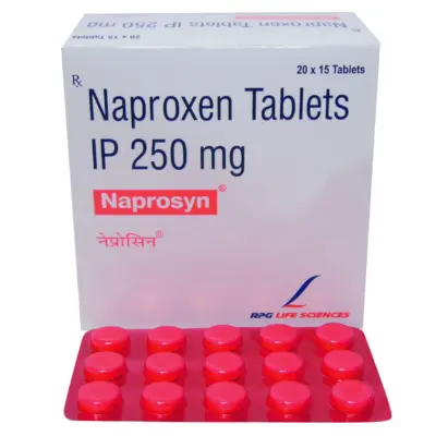
Oral NSAIDs: Medications such as Celecoxib, Naproxen, or Ibuprofen are utilized for symptomatic flares. They must be prescribed at the lowest effective dose for the shortest duration necessary, often paired with a protective Proton Pump Inhibitor (PPI) in individuals with past stomach ulcers or elevated gastrointestinal risk.
-
Intra-Articular Joint Injections:
-
Corticosteroid Injections (e.g., Triamcinolone): Injected directly into the joint space to provide rapid anti-inflammatory pain relief during acute flares, typically lasting 4 to 12 weeks. Frequent, repeated injections should be avoided, as multiple corticosteroid injections over short intervals can accelerate cartilage loss.
-
Hyaluronic Acid Viscosupplementation: Supplementing natural joint fluid with injected hyaluronic acid to improve joint lubrication and shock absorption in mild-to-moderate knee Osteoarthritis.
2. Disease-Modifying Pharmacotherapy for Autoimmune Arthritis (RA, PsA, SpA)
For inflammatory and autoimmune arthropathies, the primary treatment strategy is Treat-to-Target (T2T)—initiating aggressive disease-modifying therapy early to halt systemic inflammation, stop bone erosions, and achieve complete clinical remission:
-
Conventional Synthetic DMARDs (csDMARDs):
-
Methotrexate: The foundational, first-line anchor medication for Rheumatoid Arthritis worldwide. Taken once weekly (never daily), it inhibits dihydrofolate reductase and promotes adenosine release, blunting the autoimmune response. It is co-prescribed with daily or weekly folic acid to prevent common side effects like oral mouth sores and nausea.
-
Leflunomide, Sulfasalazine, and Hydroxychloroquine: Frequently utilized as alternative monotherapies or combined with methotrexate (e.g., “Triple Therapy”) in resistant disease.
-
Targeted Biologic DMARDs (bDMARDs): Advanced injectable or infusional medications engineered to block specific inflammatory cytokines driving joint damage:
-
Anti-TNF Biologics (Infliximab, Adalimumab, Etanercept): Neutralize Tumor Necrosis Factor-alpha.
-
IL-6 Receptor Antagonists (Tocilizumab, Sarilumab): Inhibit Interleukin-6 signaling, reducing systemic inflammation and CRP levels.
-
T-Cell Costimulation Blockers (Abatacept): Prevents full activation of autoreactive T-lymphocytes.
-
B-Cell Depleting Monoclonal Antibodies (Rituximab): Targets CD20-positive B-cells, clearing autoantibody production.
-
Pre-Treatment Safety Check: Every patient must undergo screening for latent tuberculosis and Hepatitis B before starting any biologic, as immune suppression can trigger dormant infections to reactivate.
-
Targeted Synthetic DMARDs (Oral JAK Inhibitors):
-
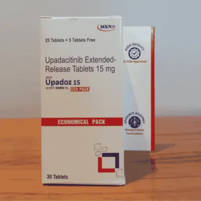
Molecules: Tofacitinib, Baricitinib, Upadacitinib.
-
Mechanism: Oral small-molecule tablets that enter cells to block Janus kinase (JAK-STAT) enzymes, shutting down multiple inflammatory cytokine signals simultaneously, providing rapid joint relief for patients who do not respond to biologics.
3. Medical Management of Gout
-
Treating Acute Flares: Rapid anti-inflammatory control using oral Colchicine (started within 24 hours of symptom onset), short-course oral corticosteroids, or high-dose NSAIDs to resolve the acute crystal attack.
-
Long-Term Urate-Lowering Therapy (ULT): Treating the root cause by keeping serum uric acid strictly below 6.0 mg/dL in patients with tophi) to dissolve existing crystal deposits and prevent new attacks:
-
Allopurinol: The primary first-line xanthine oxidase inhibitor, started at low doses (100 mg daily) and titrated upward based on monthly serum uric acid tests.
-
Febuxostat: An alternative xanthine oxidase inhibitor for patients with allopurinol intolerance or significant renal impairment.
4. Orthopedic Surgical Interventions
When conservative and medical therapies no longer relieve pain, or when structural joint destruction causes severe functional disability:
-
Total Joint Arthroplasty (Joint Replacement): Surgical removal of the damaged, arthritic cartilage and subchondral bone, replaced with precision prosthetic implants made of medical-grade titanium, cobalt-chromium alloys, and high-density polyethylene. Total Knee Arthroplasty (TKA) and Total Hip Arthroplasty (THA) are among the most successful procedures in modern surgery, providing lasting pain relief and restoring physical independence.
-
Joint-Preserving Surgeries (Osteotomy): Realignment of bones around the knee (such as high tibial osteotomy) to shift weight-bearing forces away from an arthritic compartment toward healthy cartilage in younger, active patients.
-
Arthrodesis (Joint Fusion): Surgical fusion of articulating bones into a single solid bone, eliminating motion to achieve pain relief in severe arthritis of the wrists, thumbs, ankles, and spine.
Physical Therapy, Joint Protection, and Everyday Movement
Non-pharmacological physical interventions are recommended across every clinical guideline for managing arthritis:
-
Low-Impact Aerobic Exercise: Maintaining joint movement preserves joint health. Moving joints stimulates synoviocytes to circulate synovial fluid, which supplies oxygen and nutrients to avascular cartilage. High-impact running can exacerbate joint pain, while low-impact activities—such as swimming, water aerobics, stationary cycling, and brisk walking—strengthen supporting muscles without placing sharp compressive forces on cartilage.
-
Quadriceps and Hip Muscle Strengthening: Strong muscles absorb mechanical shock before it reaches the joint. In knee Osteoarthritis, targeted physical therapy focused on strengthening the quadriceps, hamstrings, and hip abductor muscles reduces joint loading forces by up to 30%, decreasing pain and improving walking endurance.
-
Weight Management: Losing 10% of total body weight in an overweight individual cuts knee osteoarthritis pain in half, reduces cartilage degradation rates, and significantly lowers systemic circulating inflammatory adipokines.
-
Joint Protection Ergonomics:
-
Use larger, stronger joints to carry loads (e.g., carrying shoulder bags or groceries using the forearms rather than clutching handles with finger joints).
-
Avoid keeping joints locked in a single fixed posture for extended periods; stretch and change positions every 30 to 45 minutes during desk work.
-
Use assistive devices—such as wide-grip utensils, jar openers, ergonomic tools, and well-fitted walking canes held in the hand opposite the affected hip or knee—to reduce localized joint stress.
Which Medical Specialist Should You Consult?
A thorough evaluation ensures that joint symptoms are correctly categorized and treated early:
-
Rheumatologist: A physician specialized in diagnosing and managing systemic autoimmune conditions, inflammatory arthropathies (Rheumatoid Arthritis, Psoriatic Arthritis, Ankylosing Spondylitis), and complex metabolic diseases like gout. Rheumatologists lead disease-modifying therapies, DMARD protocols, and biologic monitoring.
-
Orthopedic Surgeon: A surgical specialist who evaluates mechanical joint degeneration, performs joint aspirations and injections, conducts arthroscopic surgeries, and performs partial or total joint replacements (arthroplasties).
-
Primary Care Physician / General Practitioner: The initial doctor to screen for joint symptoms, order baseline blood counts, evaluate cardiovascular risks, prescribe first-line pain management, and coordinate specialist referrals.
-
Physical Therapist (PT): Develops structured, individualized exercise regimens to build muscle strength around affected joints, correct gait mechanics, and improve range of motion.
-
Occupational Therapist (OT): Helps individuals adapt everyday home and work environments, providing specialized hand splints and recommending assistive devices to protect delicate finger and wrist joints.
Frequently Asked Questions
Can arthritis be cured completely?
Most forms of chronic arthritis—such as Osteoarthritis and Rheumatoid Arthritis—do not have a permanent medical cure, but they can be managed effectively. In autoimmune arthritis (RA and PsA), modern disease-modifying medications (DMARDs and biologics) can induce complete clinical remission, stopping pain, preventing bone erosion, and allowing individuals to lead active, normal lives. In Gout, keeping uric acid low with medications like Allopurinol can dissolve all urate crystals, curing the patient of future acute attacks. In severe localized Osteoarthritis, replacing the damaged joint with a prosthetic implant (arthroplasty) removes the arthritic joint surface and eliminates the pain.
What is the primary difference between Osteoarthritis and Rheumatoid Arthritis?
The fundamental difference lies in their underlying causes. Osteoarthritis is primarily a localized degenerative condition where mechanical wear, low-grade inflammation, and age cause the smooth cartilage covering the ends of bones to break down. Rheumatoid Arthritis is an autoimmune disease where the body’s immune system mistakenly attacks the synovial lining of joints throughout the body, causing warm, swollen, symmetrical joint inflammation that can erode bone and cause systemic complications like fatigue and fever.
Does cold, damp weather really make arthritis pain worse?
Yes, many patients report that cold, damp weather or rainy days increase joint stiffness and aching. While weather changes do not cause arthritis or worsen structural joint damage, scientific studies suggest that drops in barometric (atmospheric) pressure allow tissues inside the joint capsule to slightly expand. In joints that are already inflamed or lacking protective cartilage, this subtle expansion can irritate sensitive nerve endings, making pain and stiffness more noticeable.
Should I rest my joints or exercise if I have arthritis?
While you should rest a joint during an acute, severe inflammatory flare or after a fresh injury, regular exercise is one of the most effective long-term treatments for chronic arthritis. Inactive, immobilized joints become stiffer, while surrounding muscles weaken, which increases the compressive forces placed directly on cartilage. Engaging in low-impact activities like swimming, cycling, and walking keeps cartilage nourished, maintains joint flexibility, and builds supportive muscle strength.
What foods should I avoid if I have arthritis?
For general arthritis, reducing ultra-processed foods, refined sugars, trans fats, and excessive red meat helps lower overall systemic inflammation. For individuals managing Gout, specific dietary adjustments are important: avoid purine-rich foods that drive up uric acid, including organ meats (liver, kidneys), shellfish, beer, hard liquor, and beverages sweetened with high-fructose corn syrup. Emphasize a Mediterranean-style diet rich in vegetables, legumes, whole grains, nuts, and olive oil.
What does “cracking” or “popping” in my joints mean?
Joint sounds (clinically termed crepitus) are very common. If joint cracking or popping occurs without any pain, swelling, or stiffness, it is typically harmless, caused by tiny nitrogen gas bubbles popping within the synovial fluid as joint pressure shifts. However, if joint crepitus is accompanied by pain, swelling, or a persistent “grating” or “sandpaper-like” sensation when bending the joint, it often indicates that protective articular cartilage has worn down, allowing rough subchondral bone surfaces to rub against each other.
Why is Methotrexate taken only once a week for Rheumatoid Arthritis?
Methotrexate for Rheumatoid Arthritis is taken as a single dose strictly once per week (either as oral tablets or a subcutaneous injection). Taking methotrexate once weekly provides immune-modulating and anti-inflammatory effects while allowing the body time to recover between doses. Taking methotrexate daily is a dangerous medication error that can cause severe bone marrow suppression, mouth ulcers, and life-threatening toxicity. Patients are also co-prescribed folic acid to protect healthy cells and reduce nausea.
What should I do if a single joint suddenly becomes red, swollen, hot, and severely painful?
A joint that suddenly becomes swollen, hot, bright red, and excruciatingly painful over a few hours requires immediate emergency medical evaluation. While this can represent an acute attack of Gout, it can also be a sign of Septic Arthritis—a bacterial infection inside the joint cavity. Septic arthritis is a medical emergency; bacteria can destroy joint cartilage within 24 to 48 hours and spread into the bloodstream (sepsis). A physician must aspirate fluid from the joint immediately to test for infection and begin treatment.
Medical Authority and Clinical Reference Links
-
American College of Rheumatology (ACR):
ACR Clinical Practice Guidelines: Management of Osteoarthritis, Rheumatoid Arthritis, and Gout.
Consensus clinical practice guidelines covering non-pharmacological therapies, DMARD escalation algorithms, and joint replacement timing:
-
European Alliance of Associations for Rheumatology (EULAR):
EULAR Recommendations for the Management of Early Rheumatoid Arthritis, Psoriatic Arthritis, and Knee Osteoarthritis.
International guidelines covering treat-to-target principles, early serological screening, and targeted biologic therapies:
-
World Health Organization (WHO) – Musculoskeletal Health:
Technical Information, Global Burden Estimates, and Prevention Strategies for Osteoarthritis and Rheumatoid Disorders:
-
Arthritis Foundation:
Clinical Compendia: Types of Arthritis, Biomarker Testing Guides, and Joint Protection Ergonomics:
-
Osteoarthritis Research Society International (OARSI):
OARSI Guidelines for the Non-Surgical Management of Knee, Hip, and Polyarticular Osteoarthritis: