Anemia Medicines: Iron Supplements, Injections & Treatment Guide
Anemia is a widespread hematologic disorder characterized by a deficiency in the number of healthy circulating red blood cells or a subnormal concentration of hemoglobin—the primary iron-rich protein responsible for transporting oxygen from the lungs to tissues throughout the body.
According to the World Health Organization (WHO), anemia affects more than 1.9 billion individuals globally, with the highest burden seen in women of reproductive age, pregnant women, young children, and individuals managing chronic medical conditions.
Because oxygen delivery is necessary for basic cellular metabolism, reduced hemoglobin concentrations trigger wide-ranging physiological effects, from chronic fatigue and cognitive fog to cardiovascular strain. Left unmanaged, severe anemia can lead to high-output heart failure, impaired maternal-fetal outcomes during pregnancy, and worsening organ dysfunction in patients with kidney or cardiac disease.
Managing anemia requires an accurate assessment of the underlying cause rather than simply taking non-specific supplements. Effective pharmacotherapy ranges from oral and intravenous iron formulations for nutritional deficiency to vitamin replacements, immunosuppressive therapies, and recombinant erythropoiesis-stimulating agents (ESAs) for complex chronic conditions.
This clinical guide provides a comprehensive overview of red blood cell production, causes and types of anemia, characteristic symptoms, diagnostic laboratory markers, major therapeutic drug classes, safe administration practices, and dietary considerations.
Key Facts at a Glance
-
Diagnostic Definition (WHO): Hemoglobin level below 12.0 g/dL in non-pregnant women, below 11.0 g/dL in pregnant women, and below 13.0 g/dL in adult men.
-
Global Burden: Affects approximately 25% of the world’s population; Iron Deficiency Anemia (IDA) accounts for over 50% of all cases.
-
Most Common Forms: Iron Deficiency Anemia, Anemia of Chronic Disease/Inflammation, Megaloblastic Anemia (Vitamin B12 and Folate deficiency), Aplastic Anemia, and Hemolytic Anemias.
-
Primary Diagnostic Tests: Complete Blood Count (CBC) with red blood cell indices (MCV, MCH, MCHC), Reticulocyte Count, Peripheral Blood Smear, Serum Ferritin, Total Iron-Binding Capacity (TIBC), and Transferrin Saturation (TSAT).
-
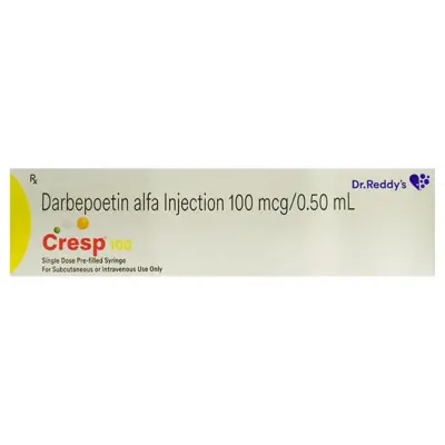
Main Medication Classes: Oral iron salts, oral iron complexes, intravenous iron formulations, Vitamin B12 (cyanocobalamin/methylcobalamin), folic acid, and Erythropoiesis-Stimulating Agents (epoetin alfa, darbepoetin alfa).
What Is Anemia? Erythropoiesis and Oxygen Transport
To understand anemia, one must examine how the body produces red blood cells through the physiological process termed erythropoiesis.
In healthy adults, red blood cells are produced continuously within the red bone marrow, predominantly in the pelvis, vertebrae, ribs, and sternum. The primary physiological driver of erythropoiesis is erythropoietin (EPO), a glycoprotein hormone synthesized and released primarily by peritubular interstitial cells in the kidneys in response to cellular hypoxia (low oxygen levels).
Once released into the bloodstream, erythropoietin binds to specific receptors on erythroid progenitor cells in the bone marrow, stimulating them to survive, multiply, and mature from proerythroblasts into reticulocytes, which are then released into the peripheral circulation to become mature erythrocytes.
Each mature red blood cell is packed with roughly 270 million hemoglobin molecules. Each hemoglobin molecule consists of four globin protein chains, each containing a central heme group holding a single ferrous iron atom. This iron atom binds reversibly to oxygen in the lungs and releases it into peripheral tissues. A mature erythrocyte circulates for approximately 100 to 120 days before being broken down by macrophages in the spleen and liver, with its iron recycled back to the bone marrow for new red blood cell synthesis.
Anemia occurs when this balance breaks down through one of three primary mechanisms:
-
Impaired or Deficient Red Blood Cell Production: Caused by a lack of essential building blocks (iron, vitamin B12, folate), insufficient bone marrow stimulation (kidney failure resulting in low erythropoietin), or bone marrow failure (aplastic anemia, myelodysplastic syndromes).
-
Blood Loss (Hemorrhage): Acute blood loss from trauma or surgery, or chronic occult blood loss from the gastrointestinal tract or heavy menstrual bleeding (menorrhagia).
-
Accelerated Red Blood Cell Destruction (Hemolysis): Premature destruction of red blood cells before their normal 120-day lifespan, driven by autoimmune antibodies, mechanical heart valves, or inherited hemoglobin abnormalities (such as sickle cell disease or thalassemia).
Major Types and Classifications of Anemia
Clinicians categorize anemia morphologically based on the physical size of the red blood cells, quantified on a Complete Blood Count by the Mean Corpuscular Volume (MCV) measured in femtoliters (fL):
1. Microcytic Anemia (MCV under 80 fL)
The red blood cells are smaller than normal, typically because the cell cannot synthesize adequate hemoglobin:
-
Iron Deficiency Anemia (IDA): The most common form worldwide. Without sufficient iron, developing erythroid cells cannot synthesize heme, leading to small (microcytic), pale (hypochromic) red blood cells.
-
Thalassemia Minor or Major: Inherited genetic mutations causing reduced or absent synthesis of normal alpha- or beta-globin chains.
-
Anemia of Chronic Disease (Early/Moderate): Systemic inflammation causes the liver to release high levels of hepcidin, a hormone that traps iron inside storage cells, preventing it from reaching the bone marrow.
-
Sideroblastic Anemia: The bone marrow produces abnormal ringed sideroblasts because it cannot properly incorporate iron into the heme molecule.
2. Normocytic Anemia (MCV between 80 and 100 fL)
The red blood cells are of normal size, but the total count or circulating concentration is low:
-
Anemia of Chronic Kidney Disease (CKD): Damaged kidneys produce insufficient erythropoietin, leading to reduced bone marrow red cell output.
-
Hemolytic Anemias: Destruction of red blood cells in the circulation, driven by autoimmune antibodies (Autoimmune Hemolytic Anemia), genetic enzyme deficiencies (G6PD deficiency), or membrane defects (hereditary spherocytosis).
-
Acute Hemorrhage: Sudden, heavy blood loss where remaining red blood cells maintain normal size and hemoglobin concentration.
-
Aplastic Anemia: Autoimmune or toxin-induced destruction of pluripotent hematopoietic stem cells in the bone marrow, resulting in low levels of all three blood cell lines (anemia, neutropenia, and thrombocytopenia).
3. Macrocytic Anemia (MCV greater than 100 fL)
The red blood cells are larger than normal, typically divided into megaloblastic and non-megaloblastic forms:
-
Megaloblastic Anemia (Vitamin B12 or Folate Deficiency): Both vitamin B12 and folate are essential cofactors for thymidine synthesis, a fundamental building block of DNA. When either nutrient is deficient, nuclear replication stalls while cytoplasmic growth continues, producing abnormally large, immature erythroblasts (megaloblasts) and oval-shaped red blood cells (macro-ovalocytes), often accompanied by hypersegmented neutrophils.
-
Non-Megaloblastic Macrocytic Anemia: Red blood cells are enlarged without defective DNA synthesis, frequently seen in chronic alcohol misuse, liver disease, untreated hypothyroidism, or treatment with specific chemotherapy drugs.
Symptoms and Warning Signs of Anemia
Symptoms of anemia depend on how low the hemoglobin level is and how quickly the condition developed. Chronic anemia that develops gradually allows compensatory mechanisms—such as increased cardiac output and shifts in the oxygen-hemoglobin dissociation curve—to minimize symptoms until hemoglobin levels drop significantly.
Common Signs of Low Tissue Oxygenation
-
Persistent Fatigue and Lethargy: Overwhelming physical and mental exhaustion that does not resolve with adequate sleep, resulting from reduced cellular oxygenation.
-
Exertional Dyspnea: Shortness of breath during routine physical activities like climbing stairs or walking short distances.
-
Skin and Mucosal Pallor: Noticeable paleness of the skin, inside of the lower eyelids (conjunctival pallor), nail beds, gums, and tongue.
-
Dizziness, Lightheadedness, and Headaches: Reduced cerebral oxygen delivery causing unsteadiness, orthostatic lightheadedness upon standing, and dull tension headaches.
-
Cold Hands and Feet: Peripheral vasoconstriction shunting oxygenated blood away from the skin toward vital central organs.
Cardiovascular Compensatory Signs
As blood carries less oxygen, the heart must pump faster and with greater force to maintain tissue oxygen delivery:
-
Tachycardia and Heart Palpitations: A rapid, pounding, or fluttering heartbeat, especially during mild exertion.
-
Flow Murmurs: Turbulent blood flow through heart valves caused by reduced blood viscosity.
-
Worsening Angina or Heart Failure: In individuals with underlying coronary artery disease, severe anemia can trigger chest pain (angina) or precipitate fluid retention and breathlessness from high-output heart failure.
Distinct Symptoms Linked to Specific Causes
-
Symptoms of Iron Deficiency:
-
Pica: An unusual craving to eat non-food substances, most commonly crushed ice (pagophagia), cornstarch, clay, or chalk.
-
Koilonychia: Brittle, flat, or spoon-shaped fingernails with raised outer edges.
-
Angular Cheilitis: Painful, cracked, red sores at the corners of the mouth.
-
Atrophic Glossitis: A smooth, swollen, sore, or burning red tongue with loss of normal surface papillae.
-
Restless Legs Syndrome (RLS): An uncomfortable, crawling urge to move the legs, particularly in the evening.
-
Symptoms of Vitamin B12 Deficiency (Neurological Involvement):
-
Peripheral Neuropathy: Symmetrical numbness, tingling, or “pins-and-needles” sensations in the toes, feet, and fingers.
-
Subacute Combined Degeneration: Loss of position and vibration sense, leading to an unsteady, clumsy gait and balance difficulties.
-
Neuropsychiatric Changes: Memory loss, depression, irritability, and cognitive decline, which can become irreversible if vitamin B12 therapy is delayed.
-
Symptoms of Hemolytic Anemia:
-
Jaundice: Yellowing of the eyes and skin resulting from high levels of unconjugated bilirubin released by broken-down red blood cells.
-
Dark or Tea-Colored Urine: High excretion of urobilinogen or hemoglobin in the urine.
-
Splenomegaly: An enlarged spleen filtering large volumes of damaged red blood cells.
Causes and Risk Factors
Anemia can result from nutritional shortages, chronic illnesses, blood loss, or genetic conditions:
1. Inadequate Intake or Malabsorption of Nutrients
-
Iron Deficiency: Insufficient intake of bioavailable dietary iron, common in strict vegan or vegetarian diets lacking supplementation.
-
Malabsorption Conditions: Celiac disease, Crohn’s disease, chronic Helicobacter pylori gastritis, or past bariatric surgery (such as gastric bypass) that bypasses or damages the duodenum and upper jejunum where iron is primarily absorbed.
-
Pernicious Anemia: An autoimmune condition where autoantibodies destroy gastric parietal cells or block intrinsic factor, a transport protein necessary for absorbing vitamin B12 in the terminal ileum.
2. Blood Loss
-
Chronic Gastrointestinal Bleeding: The leading cause of iron deficiency anemia in adult men and postmenopausal women. Common sources include peptic ulcers, colon polyps, colorectal cancer, erosive gastritis from regular NSAID use, and hemorrhoids.
-
Gynecological Blood Loss: Heavy, prolonged menstrual bleeding (menorrhagia) or uterine fibroids in premenopausal women.
-
Increased Demand During Pregnancy: Blood plasma volume expands by roughly 50% during pregnancy, requiring substantial amounts of iron and folate to support placental growth and fetal development.
3. Chronic Inflammation and Renal Disease
-
Chronic Kidney Disease (CKD): Declining kidney function reduces the production of natural erythropoietin.
-
Anemia of Chronic Disease: Driven by chronic infections (tuberculosis, HIV), autoimmune diseases (rheumatoid arthritis, lupus), or cancer. High levels of inflammatory cytokines (such as Interleukin-6) stimulate the liver to overproduce hepcidin. Hepcidin degrades ferroportin channels, trapping iron inside macrophages and liver cells, preventing it from reaching the bone marrow despite normal total body iron stores.
How Anemia Is Diagnosed: Laboratory Testing
Accurate diagnosis requires identifying the specific type and underlying cause through structured blood testing:
-
Complete Blood Count (CBC):
-
Hemoglobin (Hb) and Hematocrit (Hct): Confirms the presence and severity of anemia.
-
Mean Corpuscular Volume (MCV): Classifies the anemia as microcytic (under 80 fL), normocytic (80 to 100 fL), or macrocytic (greater than 100 fL).
-
Red Cell Distribution Width (RDW): Measures variation in red blood cell size. High RDW indicates mixed cell populations typical of early iron or nutritional deficiency.
-
Reticulocyte Count: Measures immature red blood cells released from the bone marrow. A low count indicates poor marrow production (iron deficiency, aplastic anemia), while a high count indicates the marrow is actively responding to blood loss or hemolysis.
-
Iron Studies Panel:
-
Serum Ferritin: The primary indicator of total body iron stores. A ferritin level below 30 ng/mL confirms iron deficiency. In the presence of systemic inflammation, ferritin can be elevated as an acute-phase reactant; in these cases, a ferritin level under 100 ng/mL accompanied by low transferrin saturation still indicates iron deficiency.
-
Serum Iron and Total Iron-Binding Capacity (TIBC): Evaluates circulating iron and the blood’s capacity to bind transferrin.
-
Transferrin Saturation (TSAT): Calculated as serum iron divided by TIBC, expressed as a percentage. A TSAT under 20% indicates insufficient iron available for active erythropoiesis.
-
Vitamin B12 and Serum/RBC Folate Levels: Evaluates macrocytic presentations. When B12 levels are borderline, elevated methylmalonic acid (MMA) and homocysteine levels confirm cellular B12 deficiency.
-
Stool Occult Blood and Endoscopy: In adult men and postmenopausal women with unexplained iron deficiency anemia, a fecal immunochemical test (FIT), upper endoscopy, and colonoscopy are recommended to identify or rule out hidden gastrointestinal bleeding or malignancy.
Major Classes of Anemia Medicines
Pharmacotherapy is targeted to correct the specific physiological deficiency or underlying disease process:
Oral iron replacement is the primary first-line treatment for uncomplicated, mild-to-moderate iron deficiency anemia:
-
Traditional Ferrous Iron Salts:
-
Ferrous Sulfate: The standard historical reference salt, typically providing 65 mg of elemental iron per 200 mg or 325 mg tablet.
-
Ferrous Fumarate: Contains a higher concentration of elemental iron (roughly 33% elemental iron by weight, providing about 66 to 100 mg elemental iron per dose).
-
Ferrous Gluconate: Contains less elemental iron (roughly 12% by weight, providing about 35 to 38 mg elemental iron), often preferred by individuals with digestive sensitivity.
-
Administration Principle: Ferrous iron is absorbed best in the acidic environment of the duodenum. Taking oral iron alongside Vitamin C (ascorbic acid) enhances absorption by keeping iron in its soluble ferrous state.
-
Modern Iron Complexes:
-
Ferric Maltol: A stable, non-ionic complex of ferric iron and maltol that prevents free iron from releasing prematurely in the stomach, reducing mucosal irritation and improving tolerance in patients with inflammatory bowel disease.
-
Iron Polysaccharide and Carbonyl Iron Formulations: Slower-releasing complexes designed to lower the incidence of common gastrointestinal side effects like nausea and constipation.
-
Alternate-Day Dosing: Recent clinical pharmacology studies confirm that taking an oral iron dose triggers an increase in the hormone hepcidin that lasts for 24 to 48 hours, which temporarily blocks further iron absorption. Taking oral iron every other day rather than multiple times daily results in equivalent or superior total iron absorption with significantly less nausea and constipation.
Intravenous iron is indicated when oral iron is poorly tolerated, ineffective, or when rapid iron repletion is clinically necessary (e.g., severe anemia in the third trimester of pregnancy, chronic kidney disease on dialysis, active inflammatory bowel disease, or continuous gastrointestinal blood loss):
3. Vitamin B12 and Folate Replacements
Used to treat megaloblastic anemia:
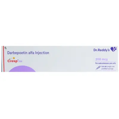
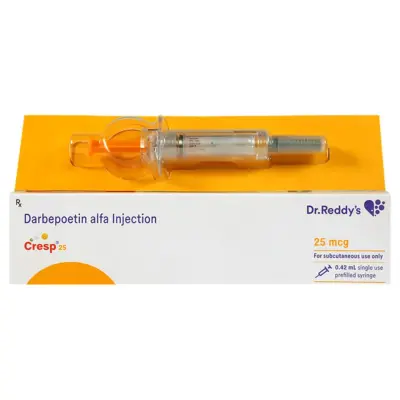
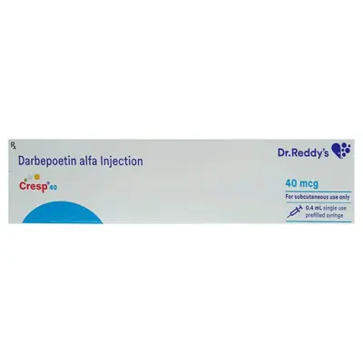
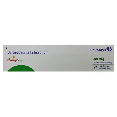
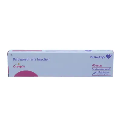
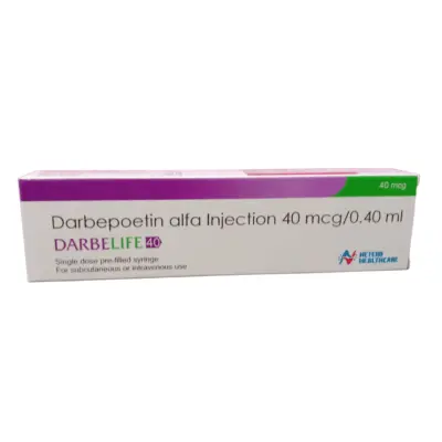
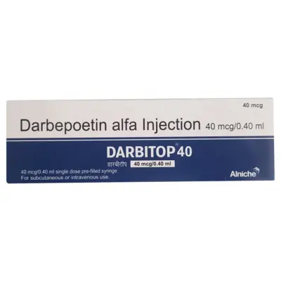
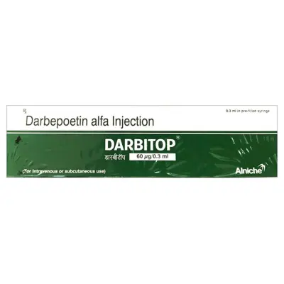
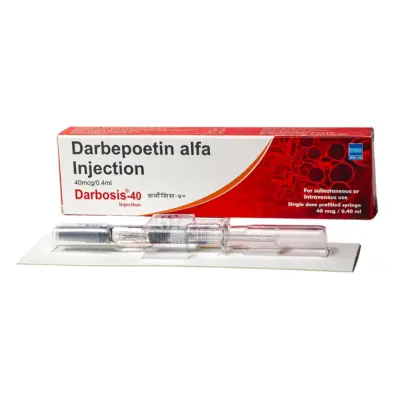
4. Erythropoiesis-Stimulating Agents (ESAs)
Recombinant biologics engineered to mimic the action of natural human erythropoietin:
5. Blood Transfusions (Packed Red Blood Cells – PRBCs)
Reserved for severe, life-threatening, or symptomatic anemia:
-
Restrictive Transfusion Strategy: Clinical guidelines recommend transfusing packed red blood cells when hemoglobin falls below 7.0 to 8.0 g/dL in stable hospitalized patients (or below 8.0 g/dL in patients with active acute coronary syndromes or undergoing major orthopedic surgery).
-
Purpose: A blood transfusion is an emergency supportive measure that rapidly stabilizes tissue oxygenation; it does not replace medical therapies targeted to fix the underlying cause of the anemia.
Safe Administration, Interactions, and Practical Use of Iron
Maximizing the effectiveness of oral iron therapy while minimizing side effects requires attention to administration and dietary timing:
Maximizing Absorption
-
Take on an Empty Stomach with Vitamin C: Oral iron salts are absorbed most efficiently when taken roughly 30 to 60 minutes before meals with water or a source of Vitamin C (such as a glass of orange juice).
-
Avoid Mineral Chelators and Dairy: Do not take oral iron with milk, yogurt, calcium supplements, or antacids. Calcium binds to iron in the digestive tract, forming insoluble complexes that block iron absorption. Separate calcium supplements or antacids by at least 2 hours.
-
Avoid Tea and Coffee: Tea and coffee contain polyphenols and tannins that bind to non-heme iron, reducing absorption by up to 60% to 70%. Avoid drinking tea or coffee within one hour before or two hours after taking an iron tablet.
Managing Common Digestive Side Effects
-
Expected Stool Changes: Oral iron turns stools a dark, greenish-black color. This is harmless and caused by unabsorbed iron passing through the digestive tract; it should not be confused with black, tarry melena from internal gastrointestinal bleeding.
-
Constipation and Nausea: If oral iron causes constipation or nausea, consider taking it every other day, taking it with a small meal, or switching to an iron complex formulation. Staying well hydrated and consuming adequate dietary fiber helps manage constipation.
-
Liquid Iron and Tooth Staining: Liquid iron drops used for children can temporarily stain tooth enamel. Dilute drops in water or fruit juice, administer them using a straw or dropper placed toward the back of the tongue, and brush teeth following the dose.
Important Drug Interactions
Oral and intravenous anemia medications interact with several commonly prescribed drugs:
-
Thyroid Hormone (Levothyroxine): Iron binds directly to levothyroxine in the stomach, preventing thyroid hormone absorption and causing TSH levels to rise. You must separate oral iron and levothyroxine by at least 4 hours.
-
Antibiotics (Fluoroquinolones and Tetracyclines): Iron forms insoluble chelates with antibiotics such as ciprofloxacin, levofloxacin, and doxycycline, impairing antibiotic absorption and risking treatment failure. Separate doses by at least 2 to 3 hours.
-
Acid-Suppressive Drugs (Proton Pump Inhibitors and H2 Blockers): Medications like omeprazole, pantoprazole, and famotidine reduce gastric acid production. Lower stomach acidity decreases the breakdown and absorption of dietary non-heme iron and vitamin B12 from foods.
Prevention and Dietary Management
While dietary adjustments alone cannot cure established, severe iron deficiency anemia once iron stores are depleted, nutrition plays a key role in maintaining healthy blood counts:
-
Heme Iron Sources: Found in animal products (such as lean poultry, fish, seafood, and eggs). Heme iron is absorbed efficiently by the body (roughly 15% to 35% absorption) and is relatively unaffected by other dietary components.
-
Non-Heme Iron Sources: Found in plant foods (such as lentils, chickpeas, beans, spinach, fortified cereals, and pumpkin seeds). Non-heme iron has a lower baseline absorption rate (2% to 10%), but its absorption is significantly enhanced when paired with Vitamin C-rich foods (bell peppers, citrus fruits, tomatoes, broccoli).
-
Dietary Folate: Found in dark green leafy vegetables (spinach, kale), legumes, asparagus, and fortified grains.
-
Dietary Vitamin B12: Naturally present in dairy products, eggs, fish, and meats. Individuals following strict vegan diets should consume fortified plant milks or take a regular daily or weekly Vitamin B12 supplement to prevent deficiency.
When to Consult a Healthcare Professional
You should seek medical evaluation if you experience:
-
Unexplained, persistent fatigue, generalized weakness, or pale skin.
-
Dizziness, lightheadedness, or feeling faint when standing up.
-
Shortness of breath or an unusually rapid heart rate during routine daily tasks.
-
Numbness, tingling, or balance changes in your hands or feet.
-
Signs of blood loss, such as black or bloody stools, heavy menstrual bleeding, or coughing up blood.
Seek immediate emergency medical care if you experience severe shortness of breath at rest, chest pain, an irregular or racing heartbeat, or sudden fainting.
Frequently Asked Questions
How long does it take for iron supplements to cure anemia?
Most patients begin feeling better, with improved energy and less shortness of breath, within 1 to 2 weeks after starting iron therapy. Laboratory tests typically show a measurable increase in hemoglobin within 2 to 4 weeks. However, it usually takes 3 to 6 months of continuous oral iron therapy to fully replenish the body’s bone marrow and liver iron stores (ferritin). Do not stop taking iron supplements as soon as your symptoms improve; continue until your doctor confirms via blood tests that your ferritin levels are restored.
Why did my doctor prescribe intravenous (IV) iron instead of pills?
Doctors recommend intravenous iron when oral iron supplements cause severe, unmanageable digestive side effects (like nausea, cramping, or constipation), when the digestive tract cannot absorb iron properly (as in celiac disease, inflammatory bowel disease, or after gastric bypass surgery), when blood loss is ongoing, or when anemia is severe and needs to be corrected quickly before surgery or in late pregnancy.
Can taking too much iron be dangerous?
Yes. Unlike many water-soluble vitamins, the human body has no active physiological pathway to excrete excess iron. Extra iron accumulates in vital organs—predominantly the liver, heart, and pancreas—where it generates oxidative stress that can lead to cirrhosis, heart failure, and diabetes (a condition known as iron overload or hemochromatosis). You should never take high-dose iron supplements unless a blood test confirms you are iron deficient and a doctor prescribes them. Always keep iron tablets locked away from children, as accidental ingestion of adult iron pills is a leading cause of fatal poisoning in young children.
What is the difference between folic acid deficiency and vitamin B12 deficiency?
Both deficiencies cause megaloblastic anemia, where red blood cells are abnormally large and fragile, resulting in identical symptoms of fatigue and pallor. However, Vitamin B12 deficiency can also damage the nervous system, leading to permanent numbness, tingling, unsteadiness, and memory problems. Folic acid deficiency does not cause nerve damage. Doctors always test both levels before treatment because taking folic acid alone can correct the blood count while allowing nerve damage from an unrecognized B12 deficiency to progress.
What does it mean if my ferritin is normal, but my hemoglobin is low?
A normal or high ferritin level alongside low hemoglobin indicates that your anemia is likely not caused by simple iron deficiency. Common causes include Anemia of Chronic Disease (where inflammation traps iron inside storage cells so it cannot be used), anemia of chronic kidney disease (lack of erythropoietin), vitamin B12 or folate deficiency, thyroid disorders, or conditions where red blood cells are broken down prematurely (hemolytic anemia). Your doctor will review other markers, such as your transferrin saturation, reticulocyte count, and kidney panels, to identify the cause.
Can drinking tea or coffee reduce iron absorption?
Yes. Both tea and coffee contain natural tannins, polyphenols, and phytates that bind to non-heme iron (the form found in plant foods and most oral supplements) in the stomach, forming insoluble compounds that your body cannot absorb. To maximize iron uptake, avoid drinking tea, coffee, or cocoa within one hour before or two hours after your iron supplement or iron-rich meals.
Why do patients with kidney disease often need erythropoietin injections?
Healthy kidneys produce about 90% of the body’s natural erythropoietin (EPO), the hormone that signals the bone marrow to manufacture new red blood cells. As chronic kidney disease progresses, the kidneys lose their ability to produce adequate EPO. Without this hormonal signal, the bone marrow produces fewer red blood cells, resulting in anemia. Synthetic erythropoiesis-stimulating agents (ESAs), such as epoetin alfa or darbepoetin alfa, replace this missing hormone to stimulate red blood cell production and reduce the need for blood transfusions.
Medical Authority and Clinical Reference Links
-
World Health Organization (WHO):
WHO Guidelines on the Prevention and Control of Anemia across the Life Course.
Public health standards on hemoglobin diagnostic cutoffs, universal screening, and nutritional iron supplementation:
-
American Society of Hematology (ASH):
ASH Clinical Practice Guidelines on the Management and Treatment of Iron Deficiency Anemia.
Authoritative guidance on oral iron dosing schedules, indications for intravenous iron, and laboratory monitoring:
-
Kidney Disease: Improving Global Outcomes (KDIGO):
KDIGO Clinical Practice Guideline for Anemia in Chronic Kidney Disease.
International consensus recommendations covering iron repletion targets (ferritin, TSAT) and the safe use of erythropoiesis-stimulating agents:
-
National Heart, Lung, and Blood Institute (NHLBI):
Clinical Health Information: Iron-Deficiency Anemia, Pernicious Anemia, and Aplastic Anemia.
Biomedical resources detailing erythropoiesis, bone marrow failure, and patient care standards:
-
American Gastroenterological Association (AGA):
AGA Clinical Practice Guidelines on the Gastrointestinal Evaluation of Iron Deficiency Anemia.
Evidence-based protocols for endoscopic and colonoscopic investigations of unexplained iron deficiency in adult populations: