Bone & Joint Health: Conditions, Treatments & Sourcing Guide
The human musculoskeletal framework consists of 206 bones connected by a network of synovial joints, articular cartilage, ligaments, tendons, and skeletal muscle. Together, these tissues provide structural support, protect vital organs, facilitate locomotion, store essential minerals like calcium and phosphate, and house the hematopoietic bone marrow responsible for blood cell production.
Throughout life, bone and joint tissues undergo continuous biological maintenance. Bones constantly balance the removal of old bone tissue with the deposition of new mineralized bone through the process of remodeling. Similarly, diarthrodial joints maintain a thin layer of articular cartilage lubricated by synovial fluid to absorb physical impact and enable frictionless movement.
When metabolic imbalances, mechanical wear, chronic inflammation, or age-related declines disrupt this equilibrium, musculoskeletal disorders develop. The most common conditions include osteoporosis (loss of bone mineral density leading to fragility fractures), osteoarthritis (breakdown of joint cartilage), inflammatory arthropathies, and osteomalacia (defective bone mineralization).
This clinical guide provides an overview of bone and joint physiology, common skeletal conditions, warning symptoms, diagnostic evaluation pathways, core pharmacological drug classes, surgical options, safe administration protocols, and preventive lifestyle strategies for the bone and joint therapies cataloged below.
Bone & Joint Clinical Matrix
-
Antiresorptive Agents (Bisphosphonates):
-
Key Molecules: Alendronate, Risedronate, Ibandronate, Zoledronic Acid.
-
Primary Mechanism: Inhibits farnesyl pyrophosphate synthase in osteoclasts, inducing osteoclast apoptosis and reducing bone breakdown.
-
Common Indications: Postmenopausal osteoporosis, male osteoporosis, glucocorticoid-induced osteoporosis, Paget’s disease of bone.
-
Critical Precautions & Monitoring: Take with plain water while sitting upright for 30 minutes; check baseline renal function (eGFR) and serum calcium; monitor for osteonecrosis of the jaw (ONJ) and atypical femur fractures.
-
Targeted RANK-Ligand Inhibitors:
-
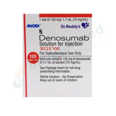
Key Molecules: Denosumab.
-
Primary Mechanism: Fully human monoclonal antibody that binds and neutralizes RANKL, preventing osteoclast formation, function, and survival.
-
Common Indications: Osteoporosis at high risk for fracture, bone loss associated with hormone ablation therapy for cancer.
-
Critical Precautions & Monitoring: Administered subcutaneously every 6 months; strict prevention of hypocalcemia (requires adequate Vitamin D and calcium intake); must not be delayed or stopped abruptly without transitioning to another antiresorptive, as cessation triggers a rebound spike in bone turnover and vertebral fracture risk.
-
Bone Anabolic (Forming) Agents:
-
Key Molecules: Teriparatide, Abaloparatide, Romosozumab.
-
Primary Mechanism: Parathyroid hormone receptor agonists (intermittent PTH signaling) stimulating osteoblast bone formation; Romosozumab binds sclerostin, driving bone formation while inhibiting resorption.
-
Common Indications: Severe osteoporosis with prior fragility fractures, very low T-scores (below -3.0), or failure of antiresorptive therapy.
-
Critical Precautions & Monitoring: Lifetime treatment limits typically capped at 18 to 24 months for PTH analogues; monitor for hypercalcemia, orthostatic hypotension, and cardiovascular risk (Romosozumab).
-
Essential Mineral & Vitamin Regimens:
-
Key Molecules: Calcium Carbonate, Calcium Citrate, Cholecalciferol (Vitamin D3), Calcitriol.
-
Primary Mechanism: Provides elemental raw mineral substrate for hydroxyapatite crystals; facilitates intestinal calcium absorption and maintains mineral homeostasis.
-
Common Indications: Osteopenia, osteoporosis prevention and co-therapy, rickets, osteomalacia, hypocalcemia.
-
Critical Precautions & Monitoring: Monitor serum calcium and 25-hydroxyvitamin D levels; avoid calcium oversupplementation to prevent nephrolithiasis (kidney stones) and vascular calcification.
-
Chondroprotective & Viscosupplementation Agents:
-
Key Molecules: Glucosamine Sulfate, Chondroitin Sulfate, Intra-Articular Hyaluronic Acid (Sodium Hyaluronate).
-
Primary Mechanism: Provides glycosaminoglycan building blocks for articular cartilage matrix; restores elastoviscosity and boundary lubrication in synovial fluid.
-
Common Indications: Mild-to-moderate knee osteoarthritis, chronic mechanical joint pain.
-
Critical Precautions & Monitoring: Check for shellfish allergies with marine-derived glucosamine; evaluate for transient localized post-injection joint swelling or effusion with intra-articular injections.
-
Anti-Inflammatory & Symptomatic Joint Analgesics:
-
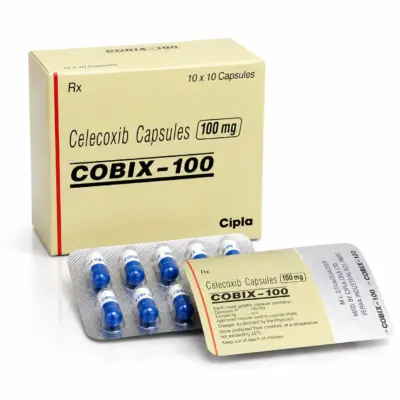
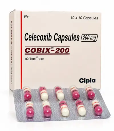
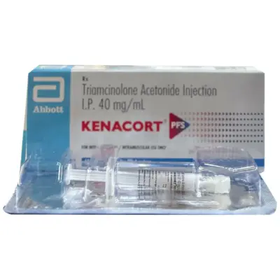
Key Molecules: Topical Diclofenac, Celecoxib, Naproxen, Intra-Articular Triamcinolone.
-
Primary Mechanism: Cyclooxygenase (COX) enzyme inhibition reducing inflammatory prostaglandins; local intra-articular anti-inflammatory suppression.
-
Common Indications: Osteoarthritis flares, acute synovitis, inflammatory joint pain.
-
Critical Precautions & Monitoring: Gastrointestinal mucosal safety (use lowest effective dose, consider co-prescribing PPIs), renal function, blood pressure monitoring; limit repeated intra-articular steroid injections to avoid accelerating cartilage thinning.
Physiology of Bone Remodeling and Synovial Joints
Musculoskeletal health relies on coordinated metabolic and mechanical processes in both bone and joint tissues:
The Bone Remodeling Cycle
Bone is a dynamic, living tissue that renews its cellular matrix throughout life. The adult skeleton replaces roughly 10% of its total mineral mass each year through the Bone Remodeling Cycle, carried out by specialized bone cells functioning within Basic Multicellular Units (BMUs):
-
Osteocytes: Mature bone cells embedded within calcified lacunae that act as mechanosensors. They detect mechanical strain and micro-damage across skeletal structures, releasing signaling molecules like sclerostin to regulate bone turnover.
-
Osteoclasts (Resorption): Multinucleated giant cells derived from hematopoietic monocyte-macrophage lineages. Stimulated by the binding of RANKL (Receptor Activator of Nuclear Factor-kappa B Ligand) to its receptor on the osteoclast surface, these cells attach to the bone surface, create a sealed acidic compartment, and secrete hydrochloric acid and cathepsin K. This dissolves the mineral component and digests the organic collagen matrix over roughly 2 to 4 weeks, carving out resorption pits (Howship’s lacunae).
-
Osteoblasts (Formation): Mononuclear cells derived from mesenchymal stem cells. They migrate into the cleared resorption cavity and synthesize osteoid—a dense, non-mineralized organic protein matrix consisting of 90% type I collagen.
-
Mineralization: Over the following 4 to 6 months, osteoblasts facilitate the crystallization of calcium and phosphate into solid hydroxyapatite crystals, mineralizing the osteoid into dense, weight-bearing lamellar bone.
Peak bone mass is achieved between ages 20 and 30. After age 40, and accelerating sharply in women following menopause due to the loss of protective estrogen, bone resorption begins to outpace bone formation. Over time, this imbalance hollows out trabecular bone architecture and thins the outer cortical shell, predisposing individuals to osteoporosis.
The Anatomy of Synovial Joints
Freely movable joints (such as the knees, hips, and shoulders) are engineered to absorb mechanical impact and facilitate smooth movement:
-
Articular Cartilage: A smooth layer of hyaline cartilage covering the bone ends. Made of chondrocytes embedded in a matrix of water, type II collagen, and proteoglycans (primarily aggrecan), it lacks direct blood vessels and nerves. It relies on physical joint movement to circulate synovial fluid, which delivers oxygen and nutrients while clearing cellular waste.
-
Synovial Membrane & Fluid: The inner lining of the joint capsule. Its synoviocytes produce synovial fluid, a clear fluid rich in hyaluronic acid and lubricin that lubricates the joint and reduces friction during movement.
-
Subchondral Bone: The bone layer immediately beneath the cartilage, designed to absorb and distribute mechanical impact into the skeletal frame.
Common Bone and Joint Conditions
Disorders affecting bones and joints fall into metabolic, degenerative, inflammatory, and traumatic categories:
-
Osteoporosis: A systemic skeletal disease characterized by low bone mass, architectural deterioration of bone tissue, and compromised structural strength, resulting in an elevated risk of fragility fractures. It is commonly termed a “silent disease” because bone loss occurs without symptoms until a fracture happens.
-
Osteopenia: A clinical classification indicating bone mineral density that is lower than normal peak density, but not yet low enough to meet the formal diagnostic threshold for osteoporosis (T-score between -1.0 and -2.5).
-
Osteomalacia & Rickets: Defective bone mineralization characterized by an accumulation of unmineralized osteoid, caused by severe Vitamin D deficiency or impaired phosphate metabolism. In adults, it leads to generalized bone pain, proximal muscle weakness, and pseudo-fractures (Looser’s zones); in children whose growth plates have not closed, it presents as rickets, causing bowing of the legs and skeletal deformities.
-
Paget’s Disease of Bone: A localized disorder of bone remodeling characterized by excessive, disorganized bone turnover. Osteoclasts resorb bone rapidly, and osteoblasts lay down replacement bone haphazardly, resulting in structurally enlarged, weak, and deformed bones prone to pain, bowing, and fractures.
2. Degenerative Joint Disorders
-
Osteoarthritis (OA): The most common joint disorder worldwide. It involves structural changes across the entire joint, including progressive breakdown of articular cartilage, thickening of subchondral bone, formation of marginal bone spurs (osteophytes), and low-grade inflammation of the synovium. It most frequently targets the knees, hips, cervical and lumbar spine, and distal finger joints.
3. Inflammatory and Autoimmune Arthropathies
-
Rheumatoid Arthritis (RA) and Spondyloarthritis: Chronic autoimmune conditions where the body’s immune system mistakenly attacks the synovial membrane or tendon insertion sites (entheses). This causes warm, swollen, painful joints that can lead to joint erosion, structural deformities, and systemic complications.
4. Traumatic and Fragility Fractures
-
Fragility Fractures: Bone fractures occurring from low-energy trauma, such as a fall from standing height or less, which would not break a healthy bone. The three classic anatomical sites for fragility fractures are:
-
The Hip (Femoral Neck and Intertrochanteric Region): Carries high risks of secondary complications, temporary or permanent loss of independent mobility, and an elevated one-year mortality rate.
-
The Spine (Vertebral Compression Fractures): Causes sudden back pain, gradual loss of height, and a hunched-forward spinal curve (kyphosis or “dowager’s hump”).
-
The Wrist (Distal Radius / Colles’ Fracture): Typically results from breaking a forward fall with an outstretched hand.
Symptoms and Warning Signs of Bone and Joint Disorders
Recognizing the clinical signs of bone and joint conditions helps ensure timely evaluation and intervention:
1. Symptoms of Joint Pathology
-
Mechanical Joint Pain: Aching pain that develops during or after movement, typically worsening with physical activity and weight-bearing, and improving with rest (classic in Osteoarthritis).
-
Joint Stiffness: Reduced joint mobility. In degenerative conditions, stiffness is brief, resolving within 15 to 30 minutes after beginning movement. In inflammatory autoimmune conditions, morning stiffness typically persists for longer than 60 minutes.
-
Crepitus: A rough, grating, crunching, or cracking sensation felt or heard when moving an affected joint, resulting from roughened cartilage surfaces or bare bone rubbing together.
-
Joint Swelling and Effusion: Fluid accumulation within the joint cavity, causing visible swelling, fullness, and restricted range of motion.
-
Joint Deformities and Instability: Progressive structural changes, such as bow-legged (varus) or knock-kneed (valgus) alignment at the knees, or bony enlargements on the fingers (Heberden’s and Bouchard’s nodes).
-
Asymptomatic Early Progression: Early osteoporosis causes no pain, warmth, or stiffness. Individuals cannot feel their bones gradually losing mineral density.
-
Sudden Severe Back Pain: Can signal an acute, spontaneous vertebral compression fracture, occurring during routine activities like bending over, coughing, or lifting light objects.
-
Gradual Height Loss: A progressive loss of height (losing 4 cm / 1.5 inches or more over several years) caused by multiple minor compression fractures collapsing anterior vertebral bodies.
-
Diffuse, Aching Bone Pain: Unlike localized joint pain, osteomalacia can cause deep, dull, widespread bone tenderness across the ribs, pelvis, and lower legs, often accompanied by proximal muscle weakness that makes climbing stairs or standing from a chair difficult.
3. Red-Flag Symptoms Requiring Urgent Evaluation
-
A single joint that becomes acutely swollen, hot, bright red, and severely painful over hours (suspected septic arthritis or acute crystal arthritis/gout).
-
Inability to bear weight on a limb following a fall.
-
New-onset severe back pain accompanied by neurological deficits, such as numbness, tingling, weakness in the legs, or loss of bowel or bladder control (suspected spinal cord or cauda equina compression).
Causes and Comprehensive Risk Factor Analysis
Bone and joint disorders result from complex interactions between non-modifiable biological factors, nutritional deficiencies, and lifestyle habits:
1. Non-Modifiable Biological Factors
-
Age: Bone mineral density peaks by age 30 and declines gradually thereafter. In joints, aging chondrocytes produce less proteoglycan, reducing cartilage elasticity and self-repair capacity.
-
Biological Sex and Estrogen Depletion: Postmenopausal women experience accelerated bone loss. Estrogen normally limits osteoclast survival and reduces RANKL expression. Following natural or surgical menopause, sharp drops in estrogen lead to accelerated osteoclast activity, with women losing up to 2% to 3% of their total bone mass annually during the first 5 to 7 years post-menopause.
-
Family History: A parental history of osteoporosis or hip fracture significantly increases an individual’s personal lifetime risk of fragility fractures.
-
Small, Thin Body Frame: Individuals with a low body mass index (BMI below 19) have less baseline bone mass and less protective soft-tissue padding around the hips, increasing fracture risk during falls.
2. Hormonal and Secondary Medical Conditions
-
Endocrine Disorders: Untreated hyperthyroidism, primary hyperparathyroidism (excess parathyroid hormone stimulating bone resorption), Cushing’s syndrome (excess cortisol), and hypogonadism (low testosterone in men).
-
Malabsorption Conditions: Celiac disease, Crohn’s disease, chronic ulcerative colitis, and bariatric surgery (such as gastric bypass) reduce intestinal absorption of calcium, magnesium, and fat-soluble Vitamin D.
-
Chronic Kidney Disease (CKD-MBD): Failing kidneys cannot activate Vitamin D into calcitriol or excrete excess phosphate, disrupting calcium homeostasis and causing secondary hyperparathyroidism and renal osteodystrophy.
3. Medication-Induced Bone and Joint Loss
-
Systemic Glucocorticoids (Steroids): The leading cause of secondary, medication-induced osteoporosis. Long-term use of oral steroids (such as prednisone at doses of 5 mg daily or higher for more than 3 months) inhibits osteoblast function, promotes osteoclast survival, decreases intestinal calcium absorption, and increases urinary calcium excretion.
-
Aromatase Inhibitors & Androgen Deprivation Therapy: Cancer therapies that block sex hormones (such as letrozole for breast cancer or leuprolide for prostate cancer) accelerate bone loss and increase fracture rates.
-
Anticonvulsants and Long-Term PPIs: Phenytoin and carbamazepine accelerate hepatic breakdown of Vitamin D, while prolonged continuous use of proton pump inhibitors can modestly reduce intestinal insoluble calcium absorption.
4. Modifiable Lifestyle Factors
-
Dietary Deficiencies: Insufficient intake of calcium, Vitamin D, and dietary protein deprives bone-forming cells of essential mineral building blocks.
-
Sedentary Lifestyle: Bones and joints require mechanical loading to stay healthy. Lack of physical activity removes the mechanical strain signals that stimulate osteoblasts to maintain bone density and reduces the circulation of lubricating fluid through joint cartilage.
-
Tobacco Smoking: Nicotine and tobacco combustion toxins impair osteoblast activity, lower circulating estrogen levels, and reduce intestinal calcium absorption.
-
Excessive Alcohol Consumption: Regular alcohol intake (more than two standard drinks daily) impairs hepatic Vitamin D hydroxylation, suppresses bone formation, and increases the physical risk of falls and traumatic fractures.
Diagnostic Evaluation: DEXA Scans, Biomarkers, and Imaging
Accurate diagnosis requires distinguishing between simple joint wear, metabolic bone mineral loss, and systemic inflammatory disease through structured testing:
Diagnostic Sequence for Bone and Joint Evaluation:
[Clinical History & Fracture Risk Assessment (FRAX)]
                         │
                         ▼
[Dual-Energy X-ray Absorptiometry (DEXA Scan: Spine & Hip)]
                         │
         ┌───────────────┴───────────────┐
         ▼                               ▼
[Metabolic Bone Evaluation]      [Articular Joint Assessment]
         │                               │
         ▼                               ▼
[Biochemical Laboratory Panels]  [Weight-Bearing X-Rays / Joint MRI]
(Serum Ca, Vit D, PTH, P1NP/CTX) (Joint Space, Osteophytes, Cartilage)
1. Dual-Energy X-ray Absorptiometry (DEXA / DXA)
The established international gold standard for measuring bone mineral density (BMD), typically evaluated at the lumbar spine and the proximal femur (total hip and femoral neck):
-
The T-Score: Compares an individual’s measured bone mineral density to the average peak bone density of a healthy young adult reference population of the same sex, expressed in standard deviations (SD):
-
Normal Bone Density: T-score of -1.0 or higher.
-
Osteopenia (Low Bone Mass): T-score between -1.0 and -2.5.
-
Osteoporosis: T-score of -2.5 or lower.
-
Severe (Established) Osteoporosis: T-score of -2.5 or lower accompanied by one or more documented low-trauma fragility fractures.
-
The Z-Score: Compares an individual’s bone density to an age-, sex-, and ethnicity-matched peer group. A Z-score of -2.0 or lower indicates abnormal bone loss that warrants investigation for secondary medical causes (e.g., hyperparathyroidism, celiac disease, or steroid toxicity).
Developed by the World Health Organization, FRAX is a computerized algorithm that integrates an individual’s femoral neck BMD with clinical risk factors (age, prior personal fracture, parental hip fracture, current smoking, systemic steroid use, rheumatoid arthritis, and alcohol intake). It calculates two prognostic scores:
-
The 10-year probability of a major osteoporotic fracture (clinical spine, forearm, hip, or shoulder).
-
The 10-year probability of a hip fracture.
-
Treatment Threshold: Clinical guidelines (such as the National Osteoporosis Foundation) recommend initiating prescription osteoporosis medications if the 10-year risk of a hip fracture is 3% or higher, or if the risk of a major osteoporotic fracture is 20% or higher, even in patients with osteopenic T-scores.
3. Biochemical Laboratory Panels
-
Mineral Homeostasis: Serum total and ionized calcium, serum phosphate, magnesium, and serum alkaline phosphatase (ALP).
-
Vitamin D Status: Serum 25-hydroxyvitamin D [25(OH)D]. Levels below 20 ng/mL represent clinical deficiency; optimal skeletal health generally targets levels between 30 and 50 ng/mL.
-
Parathyroid Hormone (PTH) and Renal Function: Baseline serum creatinine and eGFR, alongside intact PTH, to rule out secondary hyperparathyroidism or renal osteodystrophy.
-
Bone Turnover Markers (BTMs):
-
Serum C-Terminal Telopeptide (CTX): A biomarker of bone resorption reflecting osteoclast activity; useful for monitoring early response and adherence to antiresorptive therapies.
-
Procollagen Type 1 N-Terminal Propeptide (P1NP): A biomarker of active bone formation reflecting osteoblast activity, used to confirm the biological response to bone-forming therapies (such as Teriparatide).
4. Joint Imaging Modalities
-
Weight-Bearing Plain Radiographs (X-Rays): Standard imaging for assessing joint architecture. Classic signs of osteoarthritis include asymmetrical joint space narrowing (reflecting cartilage loss), subchondral bone sclerosis (thickening), subchondral bone cysts, and marginal osteophytes (bone spurs).
-
Magnetic Resonance Imaging (MRI): Visualizes soft-tissue structures, identifying early cartilage defects, subchondral bone marrow lesions/edema, meniscal tears, ligament injuries, and joint effusions before changes appear on plain X-rays.
Pharmacotherapy for Bone and Joint Health
Modern medical therapy is targeted to correct underlying metabolic deficiencies, halt bone loss, stimulate new bone formation, or protect joint structures:
1. Antiresorptive Therapies for Osteoporosis
Antiresorptive agents slow the rate of bone remodeling by inhibiting osteoclasts, allowing osteoblasts to rebuild and re-mineralize the bone matrix:
-
Oral Bisphosphonates (Alendronate, Risedronate, Ibandronate): Taken weekly or monthly. They bind firmly to exposed hydroxyapatite crystals on the bone surface. When osteoclasts absorb bone, they ingest the drug, which induces osteoclast apoptosis and decreases bone breakdown.
-
Intravenous Bisphosphonates (Zoledronic Acid): Administered as a once-yearly, 15-minute intravenous infusion (5 mg). Bypasses the digestive tract entirely, ensuring 100% adherence and providing bone protection for patients who cannot tolerate oral bisphosphonates.
-
RANK-Ligand Inhibitors (Denosumab): A subcutaneous injection administered once every 6 months. Denosumab blocks RANKL, preventing osteoclast formation and bone resorption.
-
The “Rebound Effect” Warning: Denosumab must never be delayed, skipped, or stopped without transitioning immediately to another antiresorptive agent (such as a bisphosphonate). Because denosumab does not incorporate into the bone mineral matrix, stopping treatment causes an acute rebound in osteoclast activity, leading to rapid bone density loss and an elevated risk of multiple vertebral fractures within 7 to 12 months of the missed dose.
Unlike antiresorptive drugs that merely preserve existing bone, anabolic agents stimulate osteoblasts to manufacture new, structural bone tissue:
-
Parathyroid Hormone Analogues (Teriparatide, Abaloparatide): Daily subcutaneous injections administered for 18 to 24 months. While continuous high levels of PTH resorb bone, intermittent daily pulses of PTH selectively stimulate osteoblasts to build new trabecular and cortical bone, significantly increasing bone mineral density.
-
Dual-Action Sclerostin Inhibitors (Romosozumab): A monoclonal antibody administered as monthly subcutaneous injections for 12 months. It binds and neutralizes sclerostin, a protein produced by osteocytes that normally inhibits bone formation. Neutralizing sclerostin activates the Wnt signaling pathway, which simultaneously stimulates bone formation by osteoblasts and slows bone resorption by osteoclasts.
3. Essential Nutritional and Vitamin Replacements
4. Chondroprotection and Joint Treatments
-
Symptomatic Slow-Acting Drugs for Osteoarthritis (SYSADOA): High-quality oral formulations of Glucosamine Sulfate and Chondroitin Sulfate provide glycosaminoglycan precursors for articular cartilage. Clinical trials demonstrate modest improvements in joint comfort and joint space preservation in knee osteoarthritis.
-
Intra-Articular Hyaluronic Acid (Viscosupplementation): Injected directly into the joint space under sterile technique to supplement thinned synovial fluid, providing boundary lubrication, shock absorption, and temporary anti-inflammatory pain relief lasting 3 to 6 months.
Safe Handling, Administration Protocols, and Patient Precautions
-
Strict Administration Rules for Oral Bisphosphonates:
-
Take the tablet first thing in the morning with a full glass of plain water (not mineral water, juice, tea, or coffee).
-
Do not eat, drink, or take other medications for at least 30 to 60 minutes afterward to allow proper intestinal absorption.
-
Remain sitting or standing upright for at least 30 to 60 minutes following ingestion. Never lie flat after taking the pill; lying down allows the tablet to reflux into the esophagus, which can cause chemical esophagitis and severe esophageal ulcerations.
-
Maintaining Proper Calcium Dosing: The body cannot absorb more than 500 to 600 mg of elemental calcium in a single dose. If your total daily supplemental calcium prescription is 1,000 mg, divide it into two 500 mg doses taken at different times during the day to optimize absorption and avoid gastrointestinal side effects.
-
Rare Long-Term Safety Considerations with Antiresorptives:
-
Osteonecrosis of the Jaw (ONJ): A rare condition characterized by exposed, non-healing necrotic jawbone, typically seen following invasive dental procedures (such as tooth extractions or dental implants) in patients receiving high-dose antiresorptive therapies. Patients should complete routine dental work before starting antiresorptives and maintain good oral hygiene.
-
Atypical Femur Fractures (AFF): Rare, low-trauma stress fractures occurring in the subtrochanteric shaft of the femur, linked to prolonged suppression of bone turnover (typically after 5 or more years of continuous bisphosphonate therapy). Patients who notice new, unexplained dull, aching pain in the groin or mid-thigh should contact their physician for femur imaging.
-
The “Drug Holiday”: To minimize long-term risks, guidelines recommend evaluating stable osteoporosis patients after 3 to 5 years of bisphosphonate therapy to determine whether they can safely take a temporary “drug holiday” while monitoring bone density and turnover markers.
Orthopedic Surgical Interventions
When conservative and medical therapies no longer relieve joint pain or restore physical mobility, surgical treatments may be indicated:
-
Total Joint Arthroplasty (Joint Replacement): Surgical removal of the damaged, arthritic cartilage and subchondral bone, replaced with precision prosthetic implants made of medical-grade titanium, cobalt-chromium alloys, and high-density polyethylene. Total Knee Arthroplasty (TKA) and Total Hip Arthroplasty (THA) provide lasting pain relief and restore physical function in severe osteoarthritis.
-
Percutaneous Vertebral Augmentation (Vertebroplasty & Kyphoplasty): Minimally invasive procedures for painful, non-healing acute vertebral compression fractures. Medical-grade bone cement (polymethylmethacrylate) is injected into the collapsed vertebral body to stabilize the fracture and relieve pain.
-
Open Reduction and Internal Fixation (ORIF): Surgical realignment of fractured bone fragments held in place using titanium plates, intramedullary rods, screws, or pins to ensure stable healing.
Physical Therapy, Weight-Bearing Exercise, and Joint Protection
Non-pharmacological physical interventions support long-term bone density and preserve joint mobility:
-
Weight-Bearing and Resistance Exercise for Bone Strength: Bone responds dynamically to mechanical strain. Mechanical loading creates microscopic electrical charges (piezoelectric effect) that signal osteocytes to stimulate bone mineral deposition:
-
Weight-Bearing Aerobic Exercise: Brisk walking, stair climbing, and light jogging stress bones against gravity, preserving hip and spine mineral density.
-
Progressive Resistance Training: Lifting weights or using elastic resistance bands pulls on bone via tendons, stimulating localized cortical and trabecular bone strengthening.
-
Balance Training and Fall Prevention: Most hip and wrist fragility fractures result directly from a fall. Incorporating balance-training exercises (such as Tai Chi, heel-to-toe walking, and single-leg stands) improves neuromuscular coordination, reducing fall risk by up to 30% to 50%.
-
Low-Impact Exercise for Joint Cartilage: In patients with established joint osteoarthritis, high-impact running can cause joint pain. Low-impact activities—such as swimming, water aerobics, and stationary cycling—circulate lubricating synovial fluid through joint cartilage without placing sharp compressive impact on the joints.
-
Home Safety Adjustments: Simple environmental changes help prevent falls and fractures:
-
Remove loose throw rugs and tuck away electrical cords.
-
Install sturdy grab bars inside showers and beside toilets.
-
Ensure hallways, staircases, and bathrooms are well illuminated with nightlights.
-
Wear supportive, non-slip footwear inside the home.
Which Medical Specialist Should You Consult?
A comprehensive medical evaluation coordinates accurate diagnostic workup, bone density scanning, and personalized treatment planning:
-
Orthopedic Surgeon: A surgical specialist who manages acute fractures, performs arthroscopic joint evaluations, realigns bone deformities, and executes total joint replacements (arthroplasties).
-
Endocrinologist: A medical specialist who diagnoses and manages complex metabolic bone disorders, secondary causes of osteoporosis, primary hyperparathyroidism, Paget’s disease, and multi-agent anabolic bone therapies.
-
Rheumatologist: A physician specializing in autoimmune and inflammatory joint diseases (such as rheumatoid arthritis and psoriatic arthritis), guiding disease-modifying therapies (DMARDs) and biologic infusions.
-
Physiatrist (Physical Medicine & Rehabilitation Specialist): A physician who designs non-surgical rehabilitation plans to restore physical mobility, improve posture, and manage chronic musculoskeletal pain.
-
Physical Therapist (PT): Develops personalized exercise programs to build muscle strength around weight-bearing joints, correct gait mechanics, and improve neuromuscular balance.
Frequently Asked Questions
What is the difference between osteoporosis and osteoarthritis?
Although both conditions affect the musculoskeletal system, they are entirely different diseases. Osteoarthritis is a degenerative joint disease characterized by the mechanical and biological breakdown of articular cartilage within joints, causing pain, stiffness, and reduced movement. Osteoporosis is a metabolic bone disease characterized by the loss of bone mineral density and bone tissue throughout the skeleton, which weakens bones and makes them prone to fragility fractures. Osteoporosis does not cause joint pain on its own, whereas osteoarthritis causes noticeable joint discomfort during movement.
Can osteoporosis be reversed completely?
While lost bone tissue cannot always be restored to the level of peak young adulthood, modern medical treatments can significantly rebuild bone density, restore microscopic bone architecture, and reduce future fracture risk by 50% to 70%. Anabolic bone-forming medications (such as Teriparatide and Romosozumab) actively stimulate osteoblasts to manufacture new bone, followed by antiresorptive medications (bisphosphonates or denosumab) to maintain that new bone density. Combined with calcium, Vitamin D, and resistance exercise, bone strength can improve significantly.
Why do women experience a rapid loss of bone density after menopause?
Estrogen plays a protective role in maintaining bone density by regulating osteoclasts (the cells that break down bone) and promoting bone remodeling balance. During and after menopause, the body’s natural estrogen production drops sharply. Without estrogen’s protective effect, osteoclasts survive longer and break down bone faster than osteoblasts can rebuild it. As a result, women can lose up to 2% to 3% of their total bone mass each year during the first 5 to 7 years following menopause.
What should I do if taking calcium supplements gives me constipation?
Constipation is a common side effect of high-dose calcium, particularly calcium carbonate. You can improve digestive tolerance by:
-
Switching to calcium citrate, which is absorbed more easily and tends to cause fewer digestive side effects.
-
Dividing your calcium intake into smaller doses (e.g., 500 mg twice daily with meals instead of a single 1,000 mg dose).
-
Increasing your daily dietary water intake and consuming more high-fiber foods.
-
Prioritizing dietary calcium sources (dairy products, leafy greens, fortified plant milks) so you can reduce the amount of supplemental calcium needed.
Is walking sufficient to prevent osteoporosis?
Walking is a healthy, low-impact weight-bearing exercise that helps preserve bone density in the hips and lower spine compared to sitting. However, optimal bone strengthening requires progressive resistance and impact loading. To stimulate osteocytes to signal new bone mineral deposition, walking should be paired with muscle-strengthening activities (such as resistance-band exercises, bodyweight squats, or light weightlifting) two to three times per week, alongside balance training to prevent falls.
How often should a person have a DEXA bone density scan?
For individuals with osteopenia or osteoporosis, or those receiving prescription bone-loss therapies, follow-up DEXA scans are typically recommended every 1 to 2 years to monitor treatment response and evaluate changes in bone density. For individuals with normal bone density and low clinical risk, routine rescreening is generally recommended every 3 to 5 years, or sooner if new clinical risk factors emerge (such as a fragility fracture or starting oral steroid therapy).
Why must I remain sitting or standing upright after taking an oral bisphosphonate?
Oral bisphosphonates (like alendronate and risedronate) can severely irritate the delicate lining of the esophagus. If you lie flat directly after swallowing the tablet, the pill can reflux or lodge in the esophagus, where it dissolves and can cause chemical esophagitis, deep erosions, and painful ulcers. Sitting upright, standing, or walking for at least 30 to 60 minutes allows gravity and normal digestive movement to carry the pill safely into the stomach.
What are the earliest symptoms of a fragility fracture?
Unlike traumatic breaks caused by severe accidents, a fragility fracture often presents with subtle signs. A vertebral compression fracture frequently presents as sudden, severe back pain that worsens when standing or walking, but eases slightly when lying flat. Other signs of undiagnosed spinal compression fractures include a progressive loss of physical height over several months, difficulty bending forward, or the gradual development of a rounded, hunched upper back (kyphosis). Any low-energy injury causing localized bone pain and swelling warrants an orthopedic X-ray.
Medical Authority and Clinical Reference Links
-
International Osteoporosis Foundation (IOF):
Global Patient Guidelines, Scientific Compendia, and FRAX Algorithms.
Clinical standards for bone mineral density interpretation, fragility fracture prevention, and rehabilitation:
-
Bone Health & Osteoporosis Foundation (BHOF, formerly NOF):
Clinician’s Guide to Prevention and Treatment of Osteoporosis.
Consensus clinical practice guidelines for DEXA screening intervals, calcium/Vitamin D targets, and antiresorptive therapies:
-
American College of Rheumatology (ACR):
Clinical Practice Guidelines for the Prevention and Treatment of Glucocorticoid-Induced Osteoporosis & Osteoarthritis Management.
Evidence-based treatment protocols covering secondary bone-loss prevention, joint-preservation therapies, and intra-articular interventions:
-
World Health Organization (WHO) – Collaborating Centre for Metabolic Bone Diseases:
Assessment of Fracture Risk and Its Application to Screening for Postmenopausal Osteoporosis.
The scientific foundation establishing T-score diagnostic categories and global epidemiology:
-
Osteoarthritis Research Society International (OARSI):
OARSI Guidelines for the Non-Surgical Management of Knee, Hip, and Polyarticular Osteoarthritis.
International recommendations on pharmacotherapy, topical NSAIDs, physical therapy, and biomechanical interventions: