Brain Cancer: Types, Symptoms, Causes, Diagnosis & Treatment
Brain cancer refers to the abnormal, unregulated proliferation of cells within the brain parenchyma, its protective meningeal coverings, the cranial nerves, or the central neuroendocrine structures such as the pituitary and pineal glands. Central Nervous System (CNS) malignancies are unique in human oncology.
Because the brain is enclosed within the rigid, unyielding bony skull (the cranium), even histologically benign or low-grade tumors can behave in a clinically malignant manner by compressing vital neural circuits, raising intracranial pressure, and causing irreversible neurological damage.
CNS tumors are categorized broadly into primary brain tumors (which originate directly within brain tissues) and secondary (metastatic) brain tumors (which originate in distant organs—such as the lung, breast, skin, or colon—and spread to the brain via the bloodstream). Secondary brain metastases outnumber primary brain cancers by nearly four to one. Among primary malignant brain neoplasms, Glioblastoma (GBM) represents the most common and aggressive form in adults.
Over recent years, the World Health Organization (WHO) has completely redefined the classification of CNS tumors. Diagnosis no longer relies strictly on what tumor cells look like under a light microscope; it now requires integrated molecular diagnostics, assessing genetic alterations such as IDH1/2 mutations, 1p/19q co-deletions, and MGMT promoter methylation. These molecular markers guide personalized treatments combining functional brain mapping, awake craniotomies, stereotactic radiosurgery, and targeted systemic regimens.
This clinical guide provides a comprehensive overview of neuroanatomy, the molecular classification of brain tumors, clinical warning signs, diagnostic algorithms, multimodal treatment protocols, and supportive neuro-oncological care.
Key Facts at a Glance
-
Anatomical System Affected: The Central Nervous System (brain, brainstem, cerebellum, spinal cord, cranial nerves, and meninges).
-
Primary vs. Secondary: Metastatic brain tumors are the most common brain tumors in adults; Glioblastoma is the most common primary malignant adult brain cancer.
-
Dominant Age Groups: Bimodal incidence; peaks in early childhood (ages 3 to 12) and again in older adults (ages 55 to 75).
-
Key Molecular Biomarkers: Isocitrate Dehydrogenase (IDH1/2) mutation status, 1p/19q chromosomal codeletion, MGMT promoter methylation, TERT promoter mutation, and ATRX loss.
-
Hallmark Physical Signs: Progressive morning headaches accompanied by nausea/vomiting, new-onset seizures in adulthood, focal motor weakness, cognitive/behavioral shifts, and visual deficits.
-
Diagnostic Gold Standard: Multiparametric Contrast-Enhanced Brain MRI (with functional MRI/MR spectroscopy) followed by stereotactic biopsy or surgical resection for molecular neuropathology.
-
Core Treatment Modalities: Maximal safe surgical resection (neuronavigation, awake mapping, 5-ALA fluorescence), Stupp protocol chemoradiation (Temozolomide), Stereotactic Radiosurgery (SRS), Tumor Treating Fields (TTFields), and targeted kinase inhibitors.
Neuroanatomy and the Pathology of Brain Tumors
The human brain is organized into anatomical and functional compartments enclosed within the skull:
-
The Cerebrum: The largest part of the brain, divided into two hemispheres and four lobes:
-
Frontal Lobes: Govern motor function, expressive speech (Broca’s area), judgment, executive planning, impulse control, and personality.
-
Temporal Lobes: Process auditory input, language comprehension (Wernicke’s area), and memory storage (hippocampus).
-
Parietal Lobes: Process somatosensory sensation, spatial orientation, handwriting, and sensory integration.
-
Occipital Lobes: Process visual input and interpretation.
-
The Cerebellum: Located beneath the cerebrum in the posterior fossa; coordinates voluntary motor movements, posture, gait, and fine-motor balance.
-
The Brainstem: Composed of the midbrain, pons, and medulla oblongata; bridges the brain to the spinal cord and houses autonomic centers that regulate respiration, cardiac rhythm, blood pressure, consciousness, and swallowing.
-
The Meninges: Three protective membranes wrapping the brain and spinal cord: the tough outer dura mater, the middle arachnoid mater, and the delicate inner pia mater. The subarachnoid space contains circulating cerebrospinal fluid (CSF).
-
The Blood-Brain Barrier (BBB): A physiological filtration barrier formed by specialized brain capillary endothelial cells connected by tight junctions, surrounded by a thick basement membrane and perivascular astrocytic end-feet. The BBB protects neural tissue from circulating toxins and pathogens, but also restricts more than 95% of standard systemic chemotherapy drugs from entering brain tissue.
Glial Cells and Primary Cellular Lineages
Primary brain tumors originate predominantly from glial cells—the non-neuronal supportive cells of the central nervous system:
-
Astrocytes: Star-shaped cells that maintain the blood-brain barrier, regulate extracellular neurotransmitters, and support metabolic homeostasis. Tumors derived from these cells are Astrocytomas (including Glioblastoma).
-
Oligodendrocytes: Cells that manufacture the protective myelin sheath insulating axons in the central nervous system. Tumors arising here are Oligodendrogliomas.
-
Ependymal Cells: Epithelial-like cells lining the ventricular cavities of the brain and the central canal of the spinal cord, responsible for circulating CSF. Tumors arising here are Ependymomas.
-
Meningothelial Cells: Cells within the arachnoid villi that can transform into Meningiomas (which are predominantly non-malignant, but can behave aggressively when high-grade).
-
Primitive Embryonal Cells: Undifferentiated neural precursor cells that give rise to high-grade childhood brain tumors like Medulloblastoma.
The WHO Molecular Classification of CNS Tumors
Historically, brain tumors were graded from Grade 1 to 4 solely based on histological features (such as cellular atypia, mitotic figures, vascular proliferation, and necrosis). The updated WHO Classification of Tumors of the Central Nervous System requires an integrated diagnosis combining microscopic appearance with specific molecular markers:
1. Isocitrate Dehydrogenase (IDH) Mutation Status
The presence or absence of a mutation in the IDH1 or IDH2 metabolic genes is the most important branching point in adult glioma classification:
-
IDH-Wildtype (Non-Mutated): Typically seen in older adults (median age 60+). Represents true Glioblastoma, WHO Grade 4. These tumors behave aggressively, divide rapidly, and carry a shorter median survival.
-
IDH-Mutant: Arises predominantly in younger adults (ages 30 to 50). These tumors progress more slowly and carry a significantly better overall prognosis than IDH-wildtype tumors.
2. Chromosomal 1p/19q Co-deletion
Determines the difference between astrocytic and oligodendroglial lineages:
-
If an IDH-mutant glioma displays complete, simultaneous loss (co-deletion) of the short arm of chromosome 1 and the long arm of chromosome 19 ($1p/19q\text{ co-deleted}$), along with a TERT promoter mutation, it is classified as an Oligodendroglioma (WHO Grade 2 or 3). These tumors are sensitive to alkylating chemotherapy (such as PCV or Temozolomide) and carry favorable long-term survival profiles.
-
If the tumor is IDH-mutant but retains its chromosomes (non-codeleted) and displays loss of ATRX and TP53 mutations, it is classified as an Astrocytoma, IDH-mutant (WHO Grade 2, 3, or 4).
O6-methylguanine-DNA methyltransferase (MGMT) is a cellular enzyme that repairs DNA damage caused by alkylating chemotherapy:
-
Unmethylated MGMT: The gene is active and constantly repairs DNA damage, making tumor cells resistant to temozolomide chemotherapy.
-
Methylated MGMT (Epigenetically Silenced): The repair gene is turned off, leaving the cancer cell unable to fix chemotherapy-induced DNA breaks. Patients with methylated MGMT derive significantly greater survival benefits from Temozolomide chemotherapy.
Symptoms and Clinical Signs of Brain Cancer
The clinical manifestations of a brain tumor depend on two physiological factors: general increases in pressure within the skull, and focal deficits related to the specific brain region where the tumor is growing:
1. Symptoms of Elevated Intracranial Pressure (ICP)
The skull is a rigid box of fixed volume containing brain tissue (80%), blood (10%), and cerebrospinal fluid (10%). According to the Monro-Kellie hypothesis, an expanding tumor mass compromises this balance, eventually driving intracranial pressure up:
-
The Classic Brain Tumor Headache: Present in roughly 50% of patients. It is characteristically worse in the early morning upon waking (because hypoventilation during sleep causes carbon dioxide accumulation, dilating cerebral blood vessels and increasing pressure). The headache worsens with coughing, sneezing, bending forward, or physical exertion.
-
Nausea and Projectile Vomiting: Often occurring alongside morning headaches, caused by direct stimulation of the emetic center in the medulla oblongata or sudden pressure spikes.
-
Papilledema: Swelling of the optic nerve head visible during an ophthalmoscopic fundoscopic examination, caused by high pressure transmitted along the optic nerve sheath; can lead to visual blurring or brief loss of vision.
-
Progressive Lethargy and Drowsiness: As intracranial pressure rises, cerebral blood flow drops, causing cognitive slowing, personality withdrawal, severe sleepiness, and potential coma if brain herniation occurs.
2. New-Onset Seizures in Adulthood
A new-onset seizure in an adult with no prior history of epilepsy is a major warning sign requiring an urgent brain MRI.
3. Focal Neurological Deficits (Location-Specific)
-
Frontal Lobe Lesions: Progressive changes in personality, apathy, loss of social inhibitions, impaired decision-making, and expressive language difficulty (Broca’s aphasia). Contralateral hemiparesis (weakness on the opposite side of the body) develops if the motor strip is involved.
-
Temporal Lobe Lesions: Memory impairment, receptive language difficulty where the patient speaks fluently but cannot comprehend spoken words (Wernicke’s aphasia), and superior quadrant visual field defects.
-
Parietal Lobe Lesions: Loss of sensory perception on the opposite side of the body, difficulty with reading and writing (alexia/agraphia), spatial disorientation, and right-left confusion.
-
Occipital Lobe Lesions: Visual field loss on the side opposite the tumor (homonymous hemianopia) or visual hallucinations.
-
Cerebellar Lesions: Unsteady, staggering gait (ataxia), loss of balance, intention tremors during reaching, and rapid, involuntary eye movements (nystagmus).
-
Brainstem Lesions: Cranial nerve palsies causing facial numbness, double vision (diplopia), drooping of the face, difficulty swallowing (dysphagia), and hoarseness.
Causes and Risk Factors
The vast majority of primary brain tumors arise sporadically with no identifiable single cause. However, several environmental, therapeutic, and genetic risk factors are recognized:
1. Ionizing Radiation Exposure
High-dose therapeutic ionizing radiation directed to the head or neck is the most established environmental cause of primary brain tumors:
-
Individuals treated during childhood with cranial radiation for leukemia, lymphoma, or childhood malignancies face an elevated risk of developing secondary meningiomas, high-grade gliomas, or nerve sheath tumors 10 to 30 years post-exposure.
-
Routine diagnostic imaging (low-dose dental X-rays, standard chest X-rays) does not carry this risk.
2. Inherited Genetic Syndromes
Approximately 5% of brain tumors are linked to inherited familial cancer syndromes:
-
Neurofibromatosis Type 1 (NF1): Caused by mutations in the NF1 gene on chromosome 17; predisposes to optic pathway gliomas, astrocytomas, and peripheral nerve neurofibromas.
-
Neurofibromatosis Type 2 (NF2): Caused by NF2 mutations on chromosome 22; characterized by bilateral vestibular schwannomas (acoustic neuromas), multiple meningiomas, and spinal ependymomas.
-
Li-Fraumeni Syndrome (TP53 Mutation): High lifetime risk of early-onset glioblastomas alongside breast cancer and soft-tissue sarcomas.
-
Tuberous Sclerosis Complex (TSC1/TSC2): Predisposes to subependymal giant cell astrocytomas (SEGA) and cortical tubers.
-
Von Hippel-Lindau (VHL) Disease: Predisposes to capillary hemangioblastomas in the cerebellum and spinal cord.
3. Immunosuppression
Profound compromise of the immune system—particularly in individuals living with advanced HIV/AIDS or recipients of solid organ transplants taking maintenance immunosuppressants—substantially increases the risk of Primary Central Nervous System Lymphoma (PCNSL), driven by Epstein-Barr Virus (EBV) activation.
Diagnostic Evaluation: Advanced Imaging and Neuropathology
Diagnosing a brain tumor requires high-resolution neuroimaging to map anatomical margins, followed by tissue acquisition for molecular testing:
Diagnostic Pathway for Suspected Brain Tumors:
[Clinical Warning Signs: Headache, New Seizure, or Focal Deficit]
                              │
                              ▼
[Multiparametric Contrast-Enhanced Brain MRI (T1, T2, FLAIR, Diffusion)]
                              │
         ┌────────────────────┴────────────────────┐
         ▼                                         ▼
[Solitary Primary Lesion Suspected]       [Multiple Lesions at Gray-White Junction]
         │                                         │
         ▼                                         ▼
[Advanced Functional Imaging]             [Screening for Systemic Metastasis]
(fMRI, DTI Tractography, Spectroscopy)    (Chest/Abdomen/Pelvis CT + Whole-Body PET)
         │
         ▼
[Neurosurgical Intervention: Awake Craniotomy OR Stereotactic Needle Biopsy]
         │
         ▼
[Integrated Neuropathology: Histology + IDH, 1p/19q, MGMT, TERT Testing]
1. Multiparametric Magnetic Resonance Imaging (MRI)
The imaging standard for brain cancer, utilizing high-field (3-Tesla) scanners with intravenous gadolinium contrast:
-
T1-Weighted Post-Contrast: Visualizes breakdown of the blood-brain barrier; aggressive tumors (Glioblastoma) enhance brightly, showing characteristic thick, irregular, ring-enhancing margins surrounding a dark, necrotic core.
-
T2-Weighted & FLAIR (Fluid-Attenuated Inversion Recovery): Visualizes vasogenic peritumoral edema and infiltrating, non-enhancing tumor cells extending into surrounding brain tissue.
-
Diffusion-Weighted Imaging (DWI): Evaluates cellular density; highly cellular tumors (such as lymphoma and medulloblastoma) restrict water diffusion.
-
Magnetic Resonance Spectroscopy (MRS): Analyzes brain tissue biochemistry non-invasively; malignant tumors typically show an elevated choline peak (reflecting rapid cell membrane turnover) and a diminished N-acetylaspartate (NAA) peak (reflecting the loss of healthy neurons).
-
Functional MRI (fMRI) & Diffusion Tensor Imaging (DTI Tractography): Maps critical eloquent brain cortex (speech and primary motor pathways) and visualizes subcortical white-matter nerve fiber tracts, allowing neurosurgeons to plan safe surgical corridors around vital structures.
2. The Role of Computed Tomography (CT)
Non-contrast head CT is widely used in emergency departments for rapid evaluation of sudden neurological symptoms, quickly ruling out acute intracranial hemorrhage, major midline shift, or hydrocephalus. It also visualizes calcifications (common in oligodendrogliomas) and adjacent skull bone erosion.
3. Surgical Tissue Biopsy and Resection
A tissue sample is mandatory to establish an integrated molecular diagnosis. A definitive cancer diagnosis cannot be established from scans alone:
-
Image-Guided Stereotactic Biopsy: For deep-seated, surgically inaccessible tumors (in the thalamus, basal ganglia, or brainstem), a needle is guided using 3D coordinates to sample tissue cores with minimal disruption to surrounding brain tissue.
-
Open Craniotomy: The neurosurgeon removes a section of the skull to access the brain, resecting as much tumor mass as safely possible while preserving healthy neurological function.
Multimodal Treatment Approaches
Treating brain cancer requires a coordinated multidisciplinary plan including neurosurgery, radiation oncology, medical neuro-oncology, and neuro-rehabilitation:
1. Neurosurgical Oncology: Maximal Safe Resection
Surgical resection provides the primary foundation of treatment for operable tumors:
-
The Concept of “Maximal Safe Resection”: The surgical goal is to remove as much of the tumor as possible while causing zero new permanent neurological deficits. Removing more than 90% to 98% of visible contrast-enhancing tumor on post-operative MRI directly correlates with improved overall survival.
-
Advanced Intraoperative Technologies:
-
Neuronavigation: Computerized optical tracking systems that act as an intraoperative GPS, linking surgical instruments to pre-acquired 3D MRI scans in real time.
-
Awake Craniotomy with Direct Cortical Mapping: For tumors located within or adjacent to “eloquent cortex” (motor strips and language centers). The patient is gently awakened during the tumor-removal phase of surgery. The neurosurgeon stimulates the brain surface with a mild electrical probe while a neuropsychologist engages the patient in counting, naming objects, or moving fingers. If stimulation causes a speech arrest or motor twitch, that brain tissue is preserved.
-
Fluorescence-Guided Surgery (5-ALA / Gliolan): The patient drinks a solution containing 5-Aminolevulinic Acid (5-ALA) before surgery. Malignant glioma cells metabolize 5-ALA into fluorescent porphyrins; under a specialized violet-blue surgical microscope, the tumor glows bright magenta-red, helping the surgeon distinguish the infiltrative tumor edge from non-fluorescent normal brain tissue.
2. Radiation Oncology
Radiation therapy kills residual microscopic infiltrative cancer cells left behind after surgery:
-
Fractionated External Beam Radiotherapy (EBRT): Standard therapy for glioblastomas, delivered via Intensity-Modulated Radiotherapy (IMRT) or Volumetric Modulated Arc Therapy (VMAT). The target volume includes the resection cavity plus a 1.5 to 2.0 cm margin of surrounding brain tissue to cover microscopic disease, delivering a total dose of 60 Gray (Gy) in 30 daily fractions over 6 weeks.
-
Stereotactic Radiosurgery (SRS – Gamma Knife / CyberKnife): Delivers single or hypofractionated high-dose radiation beams focused with sub-millimeter precision directly onto small, discrete targets, with radiation falling off sharply to spare adjacent brain tissue. The primary treatment of choice for brain metastases, recurrent localized tumors, and vestibular schwannomas.
-
Proton Beam Therapy: Uses heavy charged proton particles that deposit their energy at a specific depth (the Bragg peak) with zero exit dose beyond the tumor, minimizing radiation exposure to healthy brain tissue in children with medulloblastoma or craniopharyngioma.
3. Systemic Chemotherapy and the Stupp Protocol
For newly diagnosed Glioblastoma, the international standard of care is the Stupp Protocol:
-
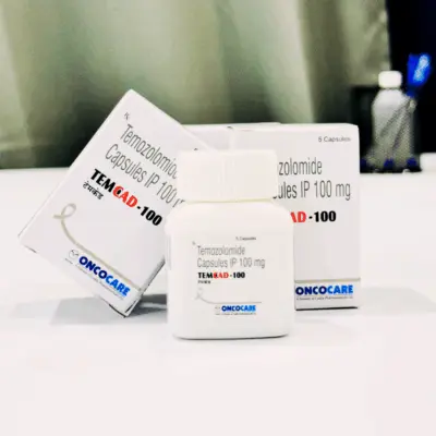
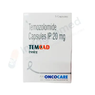
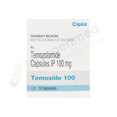
Phase 1 (Concurrent Chemoradiation): Patients receive 60 Gy of radiation over 6 weeks combined with daily oral Temozolomide (TMZ) at $75\text{ mg/m}^2$ daily, taken 7 days a week from the first to the last day of radiation.
-
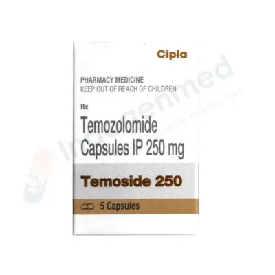
Phase 2 (Adjuvant Chemotherapy): After a 4-week break, patients receive 6 cycles of adjuvant oral Temozolomide ($150\text{ to }200\text{ mg/m}^2$ daily) taken for 5 consecutive days every 28-day cycle.
-
Mechanism of Temozolomide: An oral alkylating agent small enough to cross the blood-brain barrier. It adds methyl groups to guanine bases in DNA (specifically O6-methylguanine), causing DNA double-strand breaks that arrest cancer cell division.
4. Tumor Treating Fields (TTFields – Optune)
An FDA-approved, non-invasive regional therapy used alongside maintenance temozolomide for newly diagnosed and recurrent glioblastoma:
-
Mechanism: Transducer arrays attached directly to the shaved scalp deliver continuous, low-intensity, intermediate-frequency (200 kHz) alternating electrical fields into the brain.
-
Action: These electrical fields disrupt the alignment of charged macromolecules (tubulin and septin) necessary for mitotic spindle assembly, causing mitotic arrest and selective cancer cell death during division.
-
Administration: Worn for at least 18 hours daily, TTFields significantly extend progression-free and overall survival in clinical trials.
5. Targeted Therapies and Anti-Angiogenic Agents
-
Bevacizumab (Anti-VEGF Therapy): Glioblastomas produce high levels of Vascular Endothelial Growth Factor (VEGF), causing abnormal, leaky blood vessels that drive brain edema. Bevacizumab is an intravenous monoclonal antibody that normalizes these vessels, reducing swelling and intracranial pressure. While it improves quality of life and reduces steroid dependence in recurrent glioblastoma, it controls swelling rather than extending overall survival.
-
BRAF Inhibitors (Dabrafenib + Trametinib): For low-grade and high-grade gliomas harboring the specific BRAF V600E mutation.
-
Targeted Therapies for Brain Metastases: Systemic targeted drugs designed with high blood-brain barrier penetration can treat brain metastases without upfront whole-brain radiation:
-
Osimertinib: For EGFR-mutated non-small cell lung cancer with brain metastases.
-
Alectinib / Lorlatinib: For ALK-rearranged lung cancer metastases.
-
Dual Immunotherapy (Nivolumab + Ipilimumab): For melanoma brain metastases.
-
Tucatinib + Trastuzumab: For HER2-positive breast cancer brain metastases.
Supportive Care, Medication Safety, and Managing Complications
Supportive medications manage neurological symptoms and prevent treatment-related complications:
-
Managing Cerebral Edema with Dexamethasone:
-
Clinical Role: High-potency corticosteroid used to reduce vasogenic edema surrounding brain tumors, relieving headaches and resolving focal deficits within 24 to 48 hours.
-
Tapering Protocol: Dexamethasone should be tapered to the lowest effective dose as soon as clinical stability or surgical decompression is achieved. Prolonged use causes severe side effects, including steroid-induced myopathy, hyperglycemia/diabetes, insomnia, cushingoid changes, and opportunistic infections.
-
Antiepileptic Drug (AED) Protocols:
-
Seizure Management: Levetiracetam (Keppra) is the preferred first-line anticonvulsant for tumor-associated seizures due to its low risk of hepatic enzyme induction and absence of interactions with chemotherapy drugs.
-
Prophylaxis Guidance: Clinical guidelines advise against routinely prescribing prophylactic antiepileptic drugs to brain tumor patients who have never experienced a seizure. Prophylaxis is indicated only around the time of craniotomy (typically tapered off within 7 days post-surgery if no seizures occur).
-
Infection Prophylaxis During Chemoradiation: Patients receiving daily temozolomide alongside radiation experience lymphocyte depletion. To prevent opportunistic Pneumocystis jirovecii pneumonia (PJP), patients must take daily or three-times-weekly prophylactic antibiotics (Trimethoprim-Sulfamethoxazole / TMP-SMX) throughout concurrent chemoradiotherapy.
-
Venous Thromboembolism (VTE) Risk: Brain tumor patients carry a high risk of deep vein thrombosis and pulmonary embolism, driven by tumor pro-coagulants and limb paresis. Any sudden leg swelling warrants Doppler ultrasound evaluation.
Rehabilitation and Survivorship
Comprehensive brain cancer care integrates physical, cognitive, and emotional rehabilitation to help patients adapt and maintain independence:
-
Physical and Occupational Therapy: Targeted physical therapy helps patients recover motor strength, retrain balance, and regain safe mobility following brain surgery or hemiparesis. Occupational therapy provides adaptive strategies for activities of daily living (dressing, cooking, bathing).
-
Speech and Language Pathology: Essential for individuals with tumors in the dominant temporal or frontal lobes, helping retrain expressive language (word-finding), comprehension, and safe swallowing.
-
Cognitive Rehabilitation: Neuropsychological evaluations identify subtle deficits in short-term memory, attention span, and executive processing. Cognitive therapists provide structured mental exercises and memory aids to support daily routines.
-
Driving Safety Regulations: In most jurisdictions, experiencing a seizure or undergoing treatment for an intracranial tumor requires a mandatory driving cessation period (typically 6 to 12 months seizure-free) to ensure the safety of the patient and the public.
Frequently Asked Questions
Can primary brain cancer spread to other organs outside the brain?
Unlike most cancers that readily metastasize through the bloodstream to distant organs, primary brain tumors (such as glioblastoma) very rarely spread outside the central nervous system. The dense blood-brain barrier and the body’s basement membranes act as physical barriers preventing brain cancer cells from entering systemic blood vessels or the peripheral lymphatic system. Instead, brain tumors cause harm by invading locally into surrounding normal brain tissue or spreading through the cerebrospinal fluid (leptomeningeal dissemination) along the spinal cord.
A primary brain tumor originates directly from cells inside the brain, such as astrocytes, oligodendrocytes, or meninges (e.g., glioblastoma or astrocytoma). A metastatic (secondary) brain tumor begins as cancer in a completely different organ—most commonly the lungs, breasts, skin (melanoma), kidneys, or colon—and travels through the bloodstream to seed inside brain tissue. Metastatic brain tumors are far more common than primary brain cancers in adults.
What is the most common early symptom of a brain tumor?
The most common early symptoms are a new or changing pattern of headaches and new-onset seizures. The classic brain tumor headache is typically worse in the early morning upon waking, throbs or aches deeply, and may be accompanied by nausea or vomiting. However, symptoms depend entirely on location; a small tumor near the motor strip may cause early finger weakness or speech stumbling, while a large tumor in the frontal lobe may cause subtle personality changes, apathy, and memory slips that family members notice before physical headaches develop.
What is the difference between a benign and a malignant brain tumor?
In most parts of the body, a benign tumor is harmless because it does not invade tissues or spread. In the brain, however, both benign and malignant tumors can be life-threatening. Because the skull is a rigid box of fixed volume, a slow-growing “benign” tumor (such as a large Grade 1 meningioma) will eventually compress the brainstem, raise intracranial pressure, and cause severe deficits if it cannot be removed. A malignant brain tumor (such as glioblastoma) is both space-occupying and biologically infiltrative, sending microscopic tendrils of cancer cells deep into surrounding healthy brain tissue, making complete surgical removal impossible.
What is an awake craniotomy, and does it hurt?
An awake craniotomy is a specialized neurosurgical operation performed to remove tumors located in or near “eloquent” brain areas responsible for speech or movement. The patient is given general anesthesia or sedation while the skull bone is opened, feeling no discomfort. The patient is then gently awakened during the tumor removal phase. The brain tissue itself contains zero sensory pain receptors and feels no pain. While the patient is awake, the surgical team asks them to speak, read cards, or move their fingers while the surgeon tests brain margins with a mild electrical probe. If stimulation causes speech to pause, the surgeon knows that tissue is vital and leaves it intact, protecting the patient from permanent disability.
What does IDH mutation status mean in brain cancer?
IDH (Isocitrate Dehydrogenase) is an enzyme involved in normal cellular energy metabolism. In brain tumors, the presence of an IDH mutation indicates that the tumor originated through a specific metabolic pathway that typically affects younger adults, grows more slowly, and responds better to treatments. An IDH-wildtype status means the gene is normal; in adult diffuse gliomas, this indicates a high-grade Glioblastoma that behaves aggressively and requires immediate, intensive chemoradiation.
What is the Stupp protocol?
The Stupp protocol is the international standard treatment framework for newly diagnosed Glioblastoma. Devised by Dr. Roger Stupp, it consists of surgical resection followed by 6 weeks of external beam radiation therapy combined with daily oral Temozolomide chemotherapy. After a four-week recovery break, the patient continues with 6 cycles of maintenance Temozolomide, taken as oral capsules for 5 days out of every 28-day cycle, often paired with Tumor Treating Fields (TTFields).
Modern treatment for brain metastases has evolved beyond whole-brain radiation, which carried risks of long-term memory loss. Today, single or multiple brain metastases are treated with Stereotactic Radiosurgery (SRS)—such as Gamma Knife or CyberKnife—which delivers concentrated radiation beams focused directly onto individual tumor nodules, sparing surrounding healthy brain tissue. In addition, many modern targeted therapies and immunotherapies are designed to cross the blood-brain barrier, allowing doctors to treat systemic cancer and brain metastases simultaneously with medications.
Medical Authority and Clinical Reference Links
-
National Comprehensive Cancer Network (NCCN):
NCCN Clinical Practice Guidelines in Oncology: Central Nervous System Cancers.
Consensus clinical practice guidelines covering molecular neuropathology, surgical resection standards, the Stupp protocol, and management of brain metastases:
-
European Association of Neuro-Oncology (EANO):
EANO Guidelines on the Diagnosis and Treatment of Diffuse Gliomas in Adults.
International clinical standards covering WHO molecular classifications, awake craniotomy mapping, and systemic targeted therapies:
-
Society for Neuro-Oncology (SNO):
Clinical Guidelines, Neuro-Oncology Compendia, and Clinical Trial Standards for CNS Malignancies:
-
World Health Organization (WHO) – International Agency for Research on Cancer (IARC):
WHO Classification of Tumours of the Central Nervous System (5th Edition).
The definitive international pathological framework defining molecular and histological classifications of brain and spinal cord neoplasms:
-
American Association of Neurological Surgeons (AANS) & Congress of Neurological Surgeons (CNS):
Joint Guidelines on the Management of Adults with Newly Diagnosed Glioblastoma and Brain Metastases: