Cervical Cancer: Symptoms, Causes, Staging, Screening & Treatment
Cervical cancer is a malignant neoplasm arising from the cells of the uterine cervix—the narrow anatomical neck connecting the lower uterus to the upper vagina. Globally, it represents the fourth most common cancer in women, accounting for more than 660,000 new diagnoses and approximately 350,000 deaths every year.
In low- and middle-income regions, including parts of South Asia and sub-Saharan Africa, it remains one of the leading causes of cancer-related mortality among women in their reproductive and productive years.
Cervical cancer is distinct from most other human malignancies because its primary cause is well understood: persistent infection with high-risk human papillomavirus (hrHPV) is responsible for more than 95% to 99% of all cases. Because cervical cancer develops through identifiable, slow-progressing pre-malignant stages over 10 to 20 years, it is almost entirely preventable through population-level HPV vaccination, routine molecular screening, and early treatment of pre-invasive lesions.
For women diagnosed with invasive disease, management has evolved considerably. Diagnostic precision relies on the updated International Federation of Gynecology and Obstetrics (FIGO) staging framework, which incorporates cross-sectional imaging (MRI and PET-CT) and pathological assessments.
Treatment ranges from fertility-sparing surgeries for young women with microinvasive disease to radical hysterectomy, image-guided brachytherapy with concurrent chemoradiotherapy, and targeted immunotherapies for recurrent or metastatic disease.
This comprehensive clinical guide details cervical anatomy, HPV viral carcinogenesis, early warning signs, screening algorithms, FIGO staging, multimodal treatments, and the World Health Organization (WHO) cervical cancer elimination strategy.
Key Facts at a Glance
-
Typical Age Demographic: Most frequently diagnosed in women aged 35 to 55; peak incidence in unscreened populations between ages 40 and 50.
-
Primary Causative Agent: Persistent infection with high-risk oncogenic Human Papillomavirus (hrHPV), with Types 16 and 18 causing over 70% of cases globally.
-
Primary Histological Subtypes: Squamous Cell Carcinoma (~80%–85%) arising from the ectocervix, and Adenocarcinoma (~15%–20%) arising from the endocervical canal.
-
Key Preventative Measures: Prophylactic HPV vaccination (protecting against high-risk strains) and regular cervical screening (HPV DNA testing, Liquid-Based Cytology).
-
Hallmark Physical Symptoms: Abnormal vaginal bleeding (postcoital, intermenstrual, or postmenopausal), watery or foul-smelling discharge, and pelvic pain.
-
Diagnostic Gold Standard: Visual colposcopy with directed punch biopsy or loop electrosurgical excision procedure (LEEP) / cold-knife conization.
-
Primary Treatment Modalities: Radical Trachelectomy (fertility-sparing), Radical Hysterectomy with Pelvic Lymphadenectomy, Concurrent Chemoradiotherapy (CCRT with Cisplatin), High-Dose-Rate (HDR) Intracavitary Brachytherapy, and Immune Checkpoint Inhibitors (Pembrolizumab).
The cervix is the lower, cylindrical portion of the uterus that projects downward into the upper vagina. Anatomically, it measures approximately 3 to 4 centimeters in length and is divided into two distinct components:
-
The Ectocervix: The outer portion of the cervix visible during a standard speculum examination. It is lined by non-keratinized stratified squamous epithelium, which is resilient and designed to withstand mechanical friction.
-
The Endocervix: The inner cervical canal that connects the external os to the uterine cavity (internal os). It is lined by a single layer of mucin-secreting simple columnar epithelium arranged into crypt-like glandular folds.
The precise anatomical line where the squamous epithelium of the ectocervix meets the columnar epithelium of the endocervix is called the Squamocolumnar Junction (SCJ).
Throughout a woman’s life—particularly during puberty, pregnancy, and periods of fluctuating estrogen—the columnar epithelium naturally rolls outward onto the ectocervix (ectropion), exposing it to the acidic environment of the vagina. In response, these columnar cells undergo squamous metaplasia, transforming into mature stratified squamous cells.
The dynamic geographical band of cervical tissue between the original SCJ and the new physiological SCJ is termed the Transformation Zone (TZ). More than 90% of all cervical precancers and invasive carcinomas originate within the Transformation Zone. The continuous cellular replication and high genomic activity of these metaplastic cells make them vulnerable to oncogenic viral integration.
Pathogenesis: High-Risk HPV and Molecular Carcinogenesis
Cervical cancer does not develop overnight; it follows a well-defined molecular pathway driven by persistent viral infection:
1. Human Papillomavirus (HPV) Infection
HPV is a double-stranded DNA virus transmitted primarily through skin-to-skin sexual contact. While more than 100 HPV genotypes exist, they are clinically categorized into:
-
Low-Risk HPV Genotypes (e.g., HPV 6 and 11): Do not cause cancer; they cause benign genital warts (condylomata acuminata) and low-grade mucosal changes that resolve on their own.
-
High-Risk Oncogenic HPV Genotypes (e.g., HPV 16, 18, 31, 33, 45, 52, 58): Cause high-grade dysplasia and invasive cancer. HPV 16 and 18 are the most aggressive, with HPV 16 driving the majority of squamous cell carcinomas and HPV 18 driving adenocarcinomas.
Most sexually active adults (roughly 80%) acquire an HPV infection at some point in their lives. In more than 90% of healthy individuals, the host immune system clears the viral infection naturally within 12 to 24 months without causing lasting cellular harm.
2. Viral Persistence and Viral Integration
In roughly 5% to 10% of women, the immune system fails to clear the virus, leading to persistent hrHPV infection. Over years, persistent viral replication can cause the circular episomal viral DNA to break and integrate directly into the host human cell genome. This integration breaks the viral E2 regulatory gene, which normally keeps two viral oncoproteins in check: E6 and E7.
3. The Oncogenic Action of E6 and E7
Unchecked expression of the viral oncoproteins E6 and E7 drives malignant transformation by disabling two major tumor suppressor proteins:
-
The E6 Oncoprotein Inactivates p53: E6 binds to the host cell’s p53 tumor suppressor protein, recruiting an ubiquitin ligase (E6AP) that marks p53 for rapid destruction. Without functional p53, the host cell cannot arrest its division cycle to repair DNA mistakes or trigger apoptosis (programmed cell death), allowing damaged cells to survive and divide.
-
The E7 Oncoprotein Inactivates Retinoblastoma (pRb): E7 binds to the hypophosphorylated form of the Retinoblastoma (pRb) protein, displacing the transcription factor E2F. Free E2F moves to the cell nucleus, driving continuous entry into the S-phase of the cell cycle. As a biological byproduct of this disruption, the cellular marker p16INK4a becomes overexpressed, serving as an important diagnostic immunohistochemical biomarker for HPV-driven malignancy.
4. Progression from CIN to Invasive Carcinoma
Under the continuous influence of E6 and E7, normal cervical epithelium develops progressively severe premalignant changes known as Cervical Intraepithelial Neoplasia (CIN):
-
CIN 1 (Low-Grade Squamous Intraepithelial Lesion – LSIL): Dysplasia confined to the lower one-third of the epithelial thickness; frequently resolves on its own as the immune system clears the virus.
-
CIN 2 (Moderate Dysplasia): Atypical cells involve up to two-thirds of the epithelial thickness.
-
CIN 3 (Severe Dysplasia / Carcinoma in Situ – HSIL): Atypical cells involve the full thickness of the epithelium without breaking through the basement membrane.
-
Invasive Carcinoma: Malignant cells breach the epithelial basement membrane, invading into the underlying cervical stroma, blood vessels, and lymphatic channels. This process typically takes 10 to 20 years, providing a wide window for detection and intervention via routine screening.
Symptoms and Warning Signs of Cervical Cancer
Pre-invasive cervical dysplasia (CIN) and microinvasive Stage I cervical cancers produce no symptoms, no pain, and no visible changes on routine visual examination. Symptoms appear only when the tumor grows larger, ulcerates the surface, invades local nerve pathways, or blocks nearby pelvic organs:
1. Early and Classic Clinical Signs
-
Postcoital Vaginal Bleeding: Bleeding that occurs during or immediately after sexual intercourse is the classic, early symptom of cervical cancer. The friable, vascular tumor mass on the cervix bleeds easily when touched.
-
Intermenstrual Bleeding (Metrorrhagia): Unexplained vaginal spotting or bleeding occurring between regular menstrual periods.
-
Postmenopausal Vaginal Bleeding: Any vaginal bleeding occurring in a woman who has gone through natural menopause is abnormal and warrants immediate clinical evaluation to rule out cervical and endometrial cancers.
-
Abnormal Vaginal Discharge: A persistent, watery, pink, pale, or blood-streaked discharge that increases over time. As larger tumors undergo central necrosis, the discharge often becomes cloudy and foul-smelling.
-
Prolonged or Heavier Menstrual Cycles (Menorrhagia): Menstrual bleeding that lasts longer or is heavier than a woman’s historical baseline.
2. Advanced and Pelvic Infiltration Signs
As cervical cancer extends beyond the cervix into the parametrial tissues, pelvic sidewalls, bladder, or rectum, symptoms reflect local nerve and organ compression:
-
Deep Pelvic and Lower Back Pain: Dull, continuous, unremitting pelvic pain or lower back pain that can radiate down the back of one or both thighs along the sciatic nerve, caused by tumor invasion of the lumbosacral nerve plexus.
-
Unilateral Lower Limb Swelling (Lymphedema): Painless swelling of an entire leg caused by tumor cells invading and blocking the external iliac and pelvic lymphatic chains.
-
Urinary Symptoms and Renal Obstruction:
-
Hematuria & Urgency: Tumor ulcerating through the bladder mucosa.
-
Ureteral Obstruction: The tumor encases the ureters as they cross beneath the uterine arteries in the broad ligament, causing hydronephrosis. Unilateral or bilateral hydronephrosis can lead to silent, progressive renal failure (uremia), which is a common cause of death in untreated advanced cervical cancer.
-
Bowel and Rectal Symptoms: Tenesmus, severe rectal pain during defecation, or hematochezia (rectal bleeding) resulting from direct posterior tumor invasion into the anterior rectal wall.
-
Fistula Formation: In very advanced Stage IVA disease, necrosis of invading tumor tissue creates abnormal connections:
-
Vesicovaginal Fistula: An opening between the bladder and vagina causing continuous, involuntary leakage of urine from the vagina.
-
Rectovaginal Fistula: An opening between the rectum and vagina resulting in the passage of fecal material and gas through the vagina.
Causes and Comprehensive Risk Factor Analysis
While persistent high-risk HPV infection is the primary cause, secondary cofactors influence whether an HPV infection clears naturally or progresses to invasive cancer:
1. The Essential Cause: High-Risk HPV
Over 99% of cervical cancers harbor high-risk HPV DNA. HPV 16 alone accounts for roughly 55% to 60% of cases, while HPV 18 accounts for 10% to 15%. Other oncogenic strains include HPV 31, 33, 35, 45, 52, and 58.
2. Immunosuppression and HIV Co-Infection
A healthy immune system clears most HPV infections. In women with compromised cell-mediated immunity—most notably women living with HIV—the risk of persistent HPV infection, rapid progression from CIN to invasive cancer, and cancer recurrence is six times higher than in HIV-negative women. Because of this high risk, cervical cancer is classified as an AIDS-defining illness. Immunosuppressive therapies following organ transplantation also increase susceptibility.
3. Tobacco Smoking
Cigarette smoking is an independent cofactor for cervical squamous cell carcinoma, doubling a woman’s lifetime risk. High concentrations of tobacco carcinogens (such as nicotine and cotinine) accumulate in cervical mucus, where they damage cellular DNA and impair local Langerhans cell immune defenses, reducing the cervix’s ability to clear active HPV infections.
4. Reproductive and Sexual Factors
-
High Parity: Women who have had three or more full-term pregnancies have a higher risk, likely due to pregnancy-related hormonal shifts (high estrogen and progesterone stimulating viral transcription) and repeated cervical trauma during childbirth that exposes the transformation zone.
-
Early Age at First Intercourse: Engaging in sexual activity before age 18 exposes the immature cervical transformation zone—which is undergoing active squamous metaplasia—to HPV at a time when epithelial barriers are vulnerable.
-
Long-Term Oral Contraceptive Use: Using combined oral contraceptive pills for more than 5 to 10 years is associated with a modest increase in risk among HPV-positive women; the risk gradually declines after stopping the pill.
Population Screening and Diagnostic Algorithms
Cervical cancer screening detects high-risk viral infections and pre-cancerous cellular changes (CIN) long before invasive cancer develops:
1. Modern Screening Modalities
-
Primary HPV DNA Testing: The most sensitive and reliable screening tool available. A sample of cervical cells collected with a soft brush is tested using molecular PCR or nucleic acid hybridization to detect high-risk HPV DNA. Primary HPV screening has a high negative predictive value: a negative test provides reassurance that high-grade lesions are unlikely to develop over the following 5 years.
-
Liquid-Based Cytology (LBC / Pap Smear): Cervical cells are rinsed into a liquid preservative vial, filtered to remove mucus and blood, and examined under a microscope by a cytologist to identify abnormal cellular architecture (such as atypical squamous cells, LSIL, or HSIL) based on the Bethesda classification system.
-
Co-Testing: Performing an HPV DNA test and a liquid-based Pap smear simultaneously from the same sample.
-
Visual Inspection with Acetic Acid (VIA): In low-resource healthcare settings lacking laboratory infrastructure, dilute (3% to 5%) acetic acid is applied to the cervix. Dysplastic cells with high nuclear protein density turn chalky white (acetowhite changes), allowing immediate visual triage and treatment with cryotherapy or thermal ablation in a single visit (“screen-and-treat”).
2. Standard Population Screening Guidelines
-
Ages 21 to 29: Cervical cytology (Pap smear) alone every 3 years. Primary HPV testing is generally not recommended before age 25 to 30 because transient, harmless HPV infections are very common in young women and clear on their own.
-
Ages 30 to 65: The preferred screening strategy is primary hrHPV testing every 5 years, or co-testing (HPV + Cytology) every 5 years, or cytology alone every 3 years.
-
Discontinuation: Screening can be safely discontinued at age 65 in women who have had regular, normal screening over the previous 10 years and no history of high-grade CIN in the past 25 years.
3. Diagnostic Colposcopy and Directed Biopsy
If a screening test is positive for high-risk HPV (especially HPV 16 or 18) or shows abnormal cytology (HSIL or persistent LSIL), the patient undergoes colposcopy:
-
The Procedure: A specialized stereoscopic binocular microscope (colposcope) magnifies the illuminated cervix 6 to 40 times. The doctor applies dilute acetic acid and Lugol’s iodine solution (Schiller’s test) to highlight abnormal areas.
-
Colposcopic Features: Pre-cancerous and cancerous lesions display distinct vascular and surface patterns, including acetowhite epithelium, punctation (stippled red capillaries), mosaicism (tile-like vascular patterns), and atypical irregular branching vessels.
-
Directed Biopsy: A small biopsy forceps takes targeted tissue bites from the most abnormal areas for histopathology.
-
Endocervical Curettage (ECC): A small curette samples cells from higher up inside the endocervical canal, particularly if the entire transformation zone cannot be visualized.
Histological Classifications of Cervical Cancer
Pathologists classify invasive cervical cancers based on microscopic cellular features and immunohistochemistry:
-
Squamous Cell Carcinoma (SCC): Accounts for roughly 80% to 85% of all cervical cancers. Arises from the squamous epithelium of the ectocervix, predominantly within the transformation zone. Subdivided into keratinizing and non-keratinizing variants.
-
Adenocarcinoma: Accounts for 15% to 20% of cases. Originates from the mucus-secreting glandular columnar cells lining the endocervical canal. Adenocarcinomas are more difficult to detect on routine visual exams and standard Pap smears because they grow hidden within the cervical canal. They carry a slightly higher risk of early lymph node metastasis.
-
Adenosquamous Carcinoma: A mixed tumor showing features of both squamous and glandular malignant components, accounting for 3% to 5% of cases.
-
Neuroendocrine Tumors (Small Cell and Large Cell Carcinoma): A rare (less than 2%), aggressive subtype that lacks typical HPV association and carries a high risk of early hematogenous metastasis, requiring systemic chemotherapy regimens similar to small cell lung cancer.
Staging of Cervical Cancer: The 2018/2024 FIGO Classification
Historically, cervical cancer was one of the few cancers staged using clinical examination alone (evaluating tumor size and pelvic spread using finger palpation, speculums, and plain X-rays), because advanced imaging was unavailable in high-burden countries.
The updated FIGO Staging System allows the use of high-resolution cross-sectional imaging (pelvic MRI, CT, and whole-body FDG PET-CT) and surgical pathology to determine the stage:
FIGO Staging Overview:
Stage I   ──> Carcinoma strictly confined to the cervix uteri
Stage II  ──> Invades beyond uterus, but NOT to lower 1/3 of vagina or pelvic wall
Stage III ──> Extends to lower 1/3 of vagina, reaches pelvic wall, causes hydronephrosis,
              OR involves regional pelvic/paraaortic lymph nodes
Stage IV  ──> Invades bladder or rectal mucosa, OR spreads to distant organs
1. Stage I: Confined to the Cervix
Stage IA: Microscopic invasive carcinoma diagnosed only by histological examination, with a maximum depth of stromal invasion of ≤ 5 mm.
- Stage IA1: Measured stromal invasion ≤ 3 mm in depth.
- Stage IA2: Measured stromal invasion > 3 mm and ≤ 5 mm in depth.
Stage IB: Clinically visible lesion or microscopic invasion > 5 mm in depth, confined to the cervix.
- Stage IB1: Invasive carcinoma with a depth of invasion > 5 mm and a greatest dimension of ≤ 2 cm.
- Stage IB2: Invasive carcinoma measuring > 2 cm and ≤ 4 cm in greatest dimension.
- Stage IB3: Invasive carcinoma measuring > 4 cm in greatest dimension.
2. Stage II: Beyond the Uterus (No Lower Vagina or Pelvic Sidewall)
-
Stage IIA: Involvement of the upper two-thirds of the vagina without parametrial invasion.
- Stage IIA1: Invasive carcinoma measuring ≤ 4 cm in greatest dimension.
- Stage IIA2: Invasive carcinoma measuring > 4 cm in greatest dimension.
-
Stage IIB: Direct invasion into the parametrial tissues (the connective tissue ligaments flanking the cervix), but not extending to the bony pelvic sidewall.
3. Stage III: Lower Vagina, Pelvic Wall, Hydronephrosis, or Lymph Nodes
-
Stage IIIA: Tumor involves the lower third of the vagina, but does not extend to the pelvic sidewall.
-
Stage IIIB: Tumor extends to the bony pelvic sidewall, and/or causes hydronephrosis or non-functioning kidney.
-
Stage IIIC: Involvement of regional lymph nodes on imaging or pathology (regardless of tumor size):
-
Stage IVA: Tumor directly invades through the wall into the mucosa of the urinary bladder or rectum, or extends outside the true pelvis.
-
Stage IVB: Spread to distant organs (such as lungs, liver, bones, or distant supraclavicular lymph nodes).
Multimodal Treatment Approaches: Early-Stage Disease
Treatment for cervical cancer is divided into two approaches: surgical management for early, operable disease, and concurrent chemoradiotherapy for locally advanced disease:
1. Fertility-Sparing Surgery
For young women with early-stage disease who wish to preserve their ability to have children:
-
Cervical Conization: Cold-knife conization or LEEP with clear margins is curative for Stage IA1 disease without lymphovascular space invasion (LVSI).
-
Radical Trachelectomy (Abdominal, Vaginal, or Minimally Invasive): Indicated for Stage IA2 to selected IB1 tumors measuring 2 cm. The surgeon removes the cervix, parametrial tissues next to the cervix, and the upper 1 to 2 cm of the vagina, along with a pelvic lymph node dissection. The body and fundus of the uterus and the ovaries are preserved, and a permanent suture (cerclage) is placed around the lower uterine segment. Women can subsequently conceive and carry pregnancies, delivering via planned Cesarean section.
2. Radical Hysterectomy (The Wertheim-Meigs Operation)
The standard surgical approach for early-stage disease (Stage IA2, IB1, IB2, and selected IIA1):
-
Surgical Extent: Unlike a simple hysterectomy (which removes only the uterus and cervix), a Type C / Radical Hysterectomy removes the uterus, cervix, parametrial ligaments bilaterally out to the pelvic sidewalls, the upper one-third of the vagina, and regional pelvic lymph nodes (obturator, external iliac, internal iliac, common iliac).
-
Ovarian Preservation: In premenopausal women with squamous cell carcinoma, normal ovaries can be safely preserved or surgically relocated (ovarian transposition / oophoropexy) out of the pelvis into the upper abdomen to prevent radiation-induced premature menopause if postoperative radiation is required.
-
Surgical Approach (The LACC Trial Impact): Following the landmark prospective LACC trial, open abdominal radical hysterectomy remains the international standard of care. Minimally invasive laparoscopic or robotic radical hysterectomies were found to have higher rates of cancer recurrence and lower overall survival, likely caused by the use of intrauterine manipulators that can disrupt tumors and spread cells into the peritoneal cavity.
Multimodal Treatment: Locally Advanced Cervical Cancer (LACC)
For patients with locally advanced tumors—Stage IB3, IIA2, IIB, III, and IVA—surgery is no longer indicated, as primary radical surgery cannot achieve clear margins and adding postoperative radiation results in high complication rates.
The standard of care is definitive Concurrent Chemoradiotherapy (CCRT) combined with High-Dose-Rate Brachytherapy:
Locally Advanced Cervical Cancer Treatment Protocol:
[External Beam Radiation Therapy (EBRT: 45–50 Gy with IMRT)]
                             +
[Weekly Concurrent Radiosensitizing Cisplatin (40 mg/m² for 5–6 Weeks)]
                             │
                             ▼
[3D Image-Guided Adaptive Brachytherapy (IGABT / HDR: 28–30 Gy to Point A)]
                             │
                             ▼
[Total Combined Dose: High Biological Equivalent Dose (EQD2 ≥ 85–90 Gy)]
1. External Beam Radiation Therapy (EBRT)
Modern radiotherapy uses Intensity-Modulated Radiation Therapy (IMRT) or Volumetric Modulated Arc Therapy (VMAT) with daily image guidance:
-
Delivers approximately 45 to 50 Gray (Gy) in 25 to 28 daily fractions over 5 weeks to the whole pelvis, encompassing the cervix, uterus, parametria, upper vagina, and regional pelvic lymph nodes (extending to paraaortic fields if Stage IIIC2 nodes are positive).
-
IMRT shapes radiation beams around critical healthy organs, reducing radiation dose to the small bowel, rectum, and bladder.
2. Concurrent Radiosensitizing Chemotherapy
-
Regimen: Cisplatin administered intravenously at a dose of 40 mg once weekly for 5 to 6 cycles during external beam radiation therapy.
-
Mechanism: At this weekly dose, cisplatin does not act primarily as a systemic killer of distant cells; rather, it functions as a radiosensitizer. It forms platinum-DNA adducts that inhibit the repair of radiation-induced DNA double-strand breaks, making cancer cells more vulnerable to radiation damage.
3. Image-Guided Adaptive Brachytherapy (IGABT)
Brachytherapy is an essential, non-negotiable component of curative treatment for cervical cancer. External beam radiation alone cannot safely deliver the high total doses needed to cure cervical tumors without causing severe damage to the bladder and rectum:
-
Technique: Applicators (such as tandem and ovoids, or tandem and ring, combined with interstitial needles for bulky tumors) are placed under anesthesia into the uterine cavity and upper vagina directly against the tumor.
-
High-Dose-Rate (HDR): A high-activity radioactive source (Iridium-192) travels through catheters inside the applicator, delivering intense, targeted radiation directly into the tumor tissue over minutes, with radiation dose falling off rapidly to spare the adjacent rectum and bladder.
-
Total Target Dose: The combined radiation plan aims for a total cumulative dose of EQD2 to the high-risk clinical target volume. Treatment must be completed within a strict total timeframe of 7 to 8 weeks from the start of EBRT; treatment delays allow tumor cells to repopulate, reducing cure rates.
For women presenting with distant metastatic disease (Stage IVB) or persistent/recurrent cancer not amenable to local pelvic therapies:
-
First-Line Chemotherapy Backbone: Combination chemotherapy pairing a platinum agent with a taxane—Cisplatin (or Carboplatin) + Paclitaxel.
-
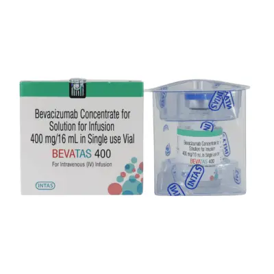
Anti-Angiogenic Therapy (Bevacizumab): Adding the anti-VEGF monoclonal antibody Bevacizumab to the chemotherapy doublet significantly improves progression-free and overall survival (as demonstrated in the GOG-240 trial) by inhibiting tumor angiogenesis.
-
Immune Checkpoint Inhibitors (Pembrolizumab): Cervical cancers express high levels of the PD-L1 ligand driven by chronic viral oncogenesis.
-
Antibody-Drug Conjugates (Tisotumab Vedotin): Approved for second-line treatment of recurrent or metastatic cervical cancer progressing after chemotherapy. It binds to Tissue Factor on cervical cancer cells, delivering the microtubule-disrupting agent MMAE directly into the cancer cell.
Safe Management and Radiation Survivorship
Cervical cancer therapies can cause predictable acute and chronic side effects that benefit from supportive care:
-
Preventing Vaginal Stenosis: Pelvic radiation therapy causes inflammation and scarring that can lead to vaginal shortening, narrowing (stenosis), and tissue dryness, which can interfere with follow-up pelvic exams and cause painful intercourse (dyspareunia).
-
Intervention: Regular use of vaginal dilators lubricated with water-soluble gel, started 2 to 4 weeks after completing brachytherapy and continued 2 to 3 times weekly for life, alongside localized vaginal moisturizers or topical estrogen creams.
-
Radiation Proctitis and Cystitis: Acute radiation can cause diarrhea, rectal urgency, and urinary frequency. Chronic radiation changes can lead to mucosal telangiectasias causing mild hematuria or rectal bleeding months or years later.
-
Management of Cisplatin-Induced Nephrotoxicity and Nausea: Patients receiving weekly cisplatin undergo vigorous intravenous pre- and post-hydration with normal saline to protect renal tubular function, alongside 5-HT3 receptor antagonists (ondansetron) and dexamethasone for nausea control.
-
Lower Limb Lymphedema: Patients who have undergone extensive pelvic lymphadenectomy or pelvic radiation should be educated on skin care, avoid cuts or infections on the legs, and use compression stockings if limb swelling occurs.
The WHO Global Elimination Strategy and Vaccination
Cervical cancer is the first human cancer that the World Health Organization has targeted for global public health elimination:
WHO 90-70-90 Elimination Targets (By 2030):
┌────────────────────────────────────────────────────────────────────────┐
│ 90% of girls fully vaccinated with the HPV vaccine by age 15           │
├────────────────────────────────────────────────────────────────────────┤
│ 70% of women screened with a high-performance test at ages 35 and 45   │
├────────────────────────────────────────────────────────────────────────┤
│ 90% of women identified with cervical disease receive treatment        │
│ (90% of pre-cancers treated; 90% of invasive cancers managed)          │
└────────────────────────────────────────────────────────────────────────┘
1. Prophylactic HPV Vaccines
Three primary recombinant virus-like particle (VLP) vaccines have been developed:
-
Bivalent Vaccine (Cervarix): Protects against high-risk types 16 and 18.
-
Quadrivalent Vaccine (Gardasil): Protects against high-risk types 16 and 18, and low-risk types 6 and 11.
-
Nonavalent Vaccine (Gardasil 9): Protects against types 6, 11, 16, 18, 31, 33, 45, 52, and 58, covering roughly 90% of all cervical-cancer-causing viral strains.
-
Dosing Schedules: Administered routinely to girls and boys aged 9 to 14 years as a 2-dose series (spaced 6 to 12 months apart) before sexual debut. Individuals aged 15 to 26 (and up to age 45 under shared clinical decision-making) receive a 3-dose series (0, 1–2, and 6 months).
-
Single-Dose Efficacy: WHO guidelines recognize that a single dose of the HPV vaccine provides comparable long-term protection against types 16 and 18, helping improve vaccine access in high-burden, low-resource countries.
Frequently Asked Questions
Can cervical cancer be prevented completely?
Yes. Cervical cancer is one of the most preventable cancers. Because nearly all cases are caused by high-risk human papillomavirus (HPV) and take 10 to 20 years to progress from pre-cancerous lesions to invasive cancer, two strategies provide protection:
-
HPV Vaccination: Vaccinating young girls and boys before they are exposed to the virus prevents over 90% of cervical cancer-causing infections.
-
Regular Screening: Routine HPV DNA testing or Pap smears detect pre-cancerous cellular changes (CIN) early, allowing doctors to treat or remove these abnormal cells before they ever become invasive cancer.
What is the most common early symptom of cervical cancer?
In its earliest stages, cervical cancer causes no symptoms at all, which is why regular screening is important. When early symptoms do emerge, the most common is abnormal vaginal bleeding, especially bleeding during or after sexual intercourse (postcoital bleeding). Other frequent early warning signs include bleeding between regular menstrual periods, new bleeding after menopause, and a watery, blood-streaked, or foul-smelling vaginal discharge.
Does having an HPV infection mean I will get cervical cancer?
No. An HPV infection does not mean you will get cancer. HPV is very common; roughly 80% of sexually active adults will contract the virus at some point in their lives. In more than 90% of people, the body’s natural immune system clears the virus within one to two years without causing harm. Only a small fraction of women who develop a persistent infection with high-risk HPV over many years are at risk of developing pre-cancerous cervical changes and invasive cancer.
What is the difference between a Pap smear and an HPV test?
Both tests use a small brush to gently collect cells from the cervix during a pelvic examination, but they look for different markers:
-
A Pap smear (cytology) examines the physical cells under a microscope to see if any cells have developed abnormal, pre-cancerous structural changes.
-
An HPV DNA test is a molecular test that checks the cells directly for the presence of genetic material from high-risk, cancer-causing types of the human papillomavirus. Modern guidelines prefer primary HPV testing because it detects cancer risk earlier and more reliably than a Pap smear alone.
Can cervical cancer be cured completely?
Yes. When cervical cancer is caught in its early stages (Stage I), cure rates exceed 85% to 90% with radical surgery or radiation therapy. For locally advanced cancers (Stage II and III) that have spread to adjacent tissues or pelvic lymph nodes, definitive concurrent chemoradiotherapy combined with image-guided brachytherapy cures roughly 60% to 70% of patients. Even in recurrent disease, modern treatments like immunotherapy and targeted drugs provide meaningful disease control.
Can a woman have children after cervical cancer treatment?
In carefully selected young women with small, early-stage tumors (Stage IA2 to small IB1 measuring 2 cm, fertility can be preserved through a specialized operation called a Radical Trachelectomy. The surgeon removes the cervix and surrounding supporting tissues while leaving the body of the uterus and the ovaries intact. Women can subsequently conceive and carry a child, delivering safely via a planned Cesarean section. For locally advanced cancers requiring chemoradiotherapy, radiation to the pelvis causes permanent ovarian failure and uterine scarring, meaning fertility preservation is not possible; these women should discuss egg or embryo freezing with a reproductive specialist before starting treatment.
What is brachytherapy, and is it necessary for cervical cancer?
Brachytherapy is an internal form of radiation therapy where small applicators are placed under anesthesia directly into the uterine cavity and vagina against the tumor. A radioactive source travels through these applicators, delivering a concentrated dose of radiation directly into the cervical tumor over minutes while sparing the adjacent bladder and rectum. Brachytherapy is a necessary component of curative treatment for locally advanced cervical cancer. External beam radiation alone cannot deliver the high radiation doses needed to eliminate cervical tumors without causing severe damage to nearby pelvic organs.
Should I still get screened if I have already received the HPV vaccine?
Yes. While HPV vaccines protect against the high-risk strains responsible for the vast majority of cervical cancers (such as types 16 and 18), they do not protect against every single cancer-causing HPV variant. In addition, women who were vaccinated after becoming sexually active may have been exposed to HPV prior to receiving the vaccine. Therefore, all vaccinated women should continue regular cervical screening according to standard national guidelines.
Medical Authority and Clinical Reference Links
-
World Health Organization (WHO) – Global Cervical Cancer Elimination Initiative:
Comprehensive Cervical Cancer Control: A Guide to Essential Practice and Global Elimination Strategy.
International standards covering HPV vaccination schedules, screening algorithms, and treatment targets:
-
International Federation of Gynecology and Obstetrics (FIGO):
FIGO Staging Classification for Cancer of the Cervix Uteri and Annual Report on Gynecological Cancers.
The primary international clinical staging framework incorporating cross-sectional imaging and pathology:
-
National Comprehensive Cancer Network (NCCN):
NCCN Clinical Practice Guidelines in Oncology: Cervical Cancer.
Consensus clinical practice guidelines for radical surgical approaches, chemoradiotherapy, brachytherapy doses, and immunotherapy:
-
American Society of Clinical Oncology (ASCO):
Secondary Prevention of Cervical Cancer: ASCO Resource-Stratified Clinical Practice Guideline.
Evidence-based global recommendations for cervical screening and management across diverse healthcare settings:
-
U.S. Preventive Services Task Force (USPSTF):
Screening for Cervical Cancer: US Preventive Services Task Force Recommendation Statement.
Evidence-based national standards for primary HPV screening intervals and cytology recommendations: