Lymphoma: Types, Symptoms, Causes, Staging & Treatment
Lymphoma is a broad group of blood cancers that originate in the lymphatic system—the vital network of vessels, nodes, and organs that forms a cornerstone of the human immune defense. Unlike solid organ carcinomas that arise in epithelial tissues (such as breast, lung, or colon cancer), lymphoma develops when lymphocytes—a specialized type of infection-fighting white blood cell—undergo malignant transformation.
These abnormal cells multiply uncontrollably, accumulate in lymph nodes and lymphoid organs, and impair the body’s natural capacity to mount an effective immune response.
Every year, more than 600,000 individuals worldwide are diagnosed with lymphoma. The disease encompasses a diverse spectrum of conditions, divided fundamentally into two major families: Hodgkin Lymphoma (HL) and Non-Hodgkin Lymphoma (NHL). Within Non-Hodgkin Lymphoma alone, modern pathology recognizes more than 70 distinct subtypes, ranging from slow-growing (indolent) conditions that can be managed over decades with active surveillance, to aggressive, fast-growing malignancies requiring immediate, intensive treatment.
Advancements in precision medicine have transformed the outlook for lymphoma patients. The integration of high-resolution PET-CT staging, targeted monoclonal antibodies (such as anti-CD20 agents), antibody-drug conjugates, bispecific T-cell engagers (BiTEs), and autologous Chimeric Antigen Receptor (CAR) T-cell therapy has substantially elevated cure and remission rates across both early and advanced stages.
This comprehensive clinical guide reviews the anatomy and physiology of the lymphatic system, the biological distinctions between Hodgkin and Non-Hodgkin disease, warning signs and “B symptoms,” underlying causes, diagnostic and staging protocols, multimodal treatment pathways, and supportive care.
Key Facts at a Glance
-
System Affected: The lymphatic system, including lymph nodes, spleen, thymus, bone marrow, tonsils, and mucosal lymphoid tissues.
-
Primary Cell Types: B-lymphocytes (accounting for roughly 85% to 90% of lymphomas), T-lymphocytes, and Natural Killer (NK) cells.
-
Major Classifications: Hodgkin Lymphoma (characterized by the microscopic presence of Reed-Sternberg cells) and Non-Hodgkin Lymphoma (which lacks Reed-Sternberg cells).
-
Age Demographics: Hodgkin Lymphoma exhibits a bimodal distribution (peaking in young adults aged 15 to 35, and again in adults over 55); Non-Hodgkin Lymphoma incidence increases steadily with age, most commonly diagnosed in individuals aged 60 and older.
-
Hallmark Physical Sign: Painless, firm, rubbery swelling of one or more lymph nodes in the neck, armpit, or groin.
-
Classic “B Symptoms”: Drenching night sweats, unexplained persistent fevers, and unintentional weight loss greater than 10% within six months.
-
Diagnostic Gold Standard: Surgical excisional lymph node biopsy with comprehensive immunohistochemistry (IHC) and molecular profiling.
-
Primary Treatments: Multi-agent immunochemotherapy (e.g., ABVD, R-CHOP), targeted monoclonal antibodies, radiation therapy, stem cell transplantation, and CAR T-cell cellular immunotherapy.
To understand lymphoma, one must examine the normal anatomy and protective function of the lymphatic system.
The lymphatic system is an extensive vascular and cellular network that works alongside the cardiovascular circulatory system. It consists of thin-walled lymphatic capillaries, larger conducting trunks, and hundreds of small, bean-shaped filtration centers known as lymph nodes. Distributed throughout the body—most densely in the neck (cervical), underarms (axillary), groin (inguinal), chest (mediastinal), and abdomen (mesenteric and retroperitoneal)—lymph nodes filter tissue fluid (lymph) to clear pathogens, cellular debris, and abnormal cells.
Key specialized organs belonging to this system include:
-
The Spleen: Located in the left upper quadrant of the abdomen; it filters blood, clears old red blood cells, stores platelets, and serves as a major reservoir of immune lymphocytes.
-
The Bone Marrow: The primary site where all blood stem cells originate and where B-lymphocytes mature.
-
The Thymus: A gland situated in the anterior mediastinum behind the sternum, responsible for the maturation and selection of T-lymphocytes during childhood.
-
Mucosa-Associated Lymphoid Tissue (MALT): Aggregates of lymphoid tissue guarding mucosal entry points throughout the body, including the tonsils, adenoids, Peyer’s patches in the small intestine, and the lining of the stomach and respiratory tree.
The Role of Normal Lymphocytes
Lymphocytes are white blood cells that coordinate adaptive immunity. They are divided into two main lineages:
-
B-Lymphocytes (B-Cells): Mature in the bone marrow and travel to lymph nodes. When exposed to a matching foreign antigen, they transform into plasma cells that manufacture targeted antibodies (immunoglobulins) to neutralize bacteria and viruses.
-
T-Lymphocytes (T-Cells): Migrate from the bone marrow to mature in the thymus gland. They differentiate into cytotoxic T-cells (which directly destroy virus-infected or abnormal cells) and helper T-cells (which release chemical cytokines to coordinate the wider immune response).
Lymphocytes must naturally undergo rapid rounds of DNA alteration and cellular division—especially inside the germinal centers of lymph nodes—to generate diverse, high-affinity antibodies. This high rate of normal genetic rearrangement makes lymphocytes particularly vulnerable to acquired DNA errors.
Lymphoma develops when sequential, non-lethal genetic lesions—such as chromosomal translocations, point mutations, or epigenetic dysregulations—occur in a developing lymphocyte. These alterations disrupt normal control points:
-
They activate cellular growth pathways (such as the B-cell receptor pathway, NF-kB signaling, or BCL-6 overexpression).
-
They turn off natural cell death signals, often through the overexpression of the anti-apoptotic protein BCL-2.
-
They block the cell from completing normal maturation.
Instead of dying after an infection clears, these abnormal lymphocytes become immortal, dividing continuously. They accumulate within lymph nodes, causing painless enlargement, and can spread through lymphatic vessels and the bloodstream to infiltrate the spleen, liver, bone marrow, and other non-lymphoid organs.
The Fundamental Divide: Hodgkin vs. Non-Hodgkin Lymphoma
Pathologists divide all lymphomas into two foundational categories based on the microscopic appearance of the tumor tissue under a microscope:
1. Hodgkin Lymphoma (HL)
Hodgkin lymphoma is defined by a unique histopathological feature: the tumor tissue consists of only a tiny minority (roughly 1% to 2%) of truly malignant cells, surrounded by an overwhelming background of non-cancerous, reactive immune cells (lymphocytes, eosinophils, neutrophils, and plasma cells).
-
The Reed-Sternberg Cell: The diagnostic hallmark of classic Hodgkin Lymphoma. These are giant, abnormal, multinucleated B-cells displaying a distinctive “owl-eye” appearance with prominent, inclusion-like nucleoli. They secrete high levels of inflammatory cytokines, which recruit the surrounding sea of non-malignant immune cells that causes the physical swelling of the lymph node.
-
Pattern of Spread: Hodgkin lymphoma is predictable in its behavior. It almost always begins in a single lymph node group (most commonly in the neck or upper chest) and spreads in an orderly, contiguous manner to the next adjacent lymph node chain before eventually moving to the spleen, liver, or bone marrow.
-
Subtypes of Hodgkin Lymphoma:
-
Classic Hodgkin Lymphoma (cHL, ~95% of cases): Divided into Nodular Sclerosis (the most common form, typical in young women, often involving the mediastinum), Mixed Cellularity (frequent in older adults and associated with Epstein-Barr virus), Lymphocyte-Rich, and Lymphocyte-Depleted.
-
Nodular Lymphocyte-Predominant Hodgkin Lymphoma (NLPHL, ~5% of cases): A rare, indolent subtype characterized by abnormal cells termed “popcorn cells” (LP cells) that express typical B-cell surface markers (CD20-positive), often behaving more like a slow-growing Non-Hodgkin lymphoma.
2. Non-Hodgkin Lymphoma (NHL)
Non-Hodgkin lymphoma encompasses all lymphomas that lack Reed-Sternberg cells. Unlike Hodgkin disease, NHL tumors are composed almost entirely of sheets of malignant lymphocytes.
-
Cellular Diversity: Approximately 85% to 90% of NHL cases arise from B-lymphocytes; the remaining 10% to 15% arise from T-lymphocytes or Natural Killer (NK) cells.
-
Pattern of Spread: Non-Hodgkin lymphoma is much less predictable. It can arise simultaneously in non-contiguous lymph node groups throughout the body and frequently involves extranodal sites—such as the stomach, intestines, skin, thyroid, brain, and bone marrow—at initial presentation.
-
Clinical Behavior Categories:
-
Indolent (Low-Grade) NHL: Slow-growing lymphomas, most commonly Follicular Lymphoma, Marginal Zone Lymphoma, and Small Lymphocytic Lymphoma (SLL). These tumors can grow quietly for years without causing noticeable symptoms. While highly responsive to initial treatments, they are traditionally challenging to eradicate permanently, often following a relapsing-remitting course over decades.
-
Aggressive (High-Grade) NHL: Rapidly dividing, symptomatic lymphomas, most commonly Diffuse Large B-Cell Lymphoma (DLBCL) and Mantle Cell Lymphoma. These tumors progress over weeks to months and are fatal if untreated, but are highly curable in a large proportion of patients using immediate, intensive immunochemotherapy.
-
Highly Aggressive NHL: Ultra-fast doubling times, such as Burkitt Lymphoma and Lymphoblastic Lymphoma, requiring immediate inpatient hospitalization, intensive combination chemotherapy, and preventive treatment for the brain and spinal cord.
Symptoms and Clinical Warning Signs of Lymphoma
The clinical manifestations of lymphoma depend on whether the disease is indolent or aggressive, which lymph node groups are enlarged, and whether extranodal organs are involved:
1. Painless Lymphadenopathy (The Primary Physical Sign)
The single most common early sign of lymphoma is persistent, non-tender swelling of one or more lymph nodes:
-
Anatomical Locations: Most frequently discovered in the neck (cervical), above the collarbone (supraclavicular), in the armpits (axillary), or in the groin (inguinal).
-
Physical Characteristics: Malignant lymph nodes are characteristically painless, firm, and rubbery, feeling distinct from the surrounding soft tissue. They are often mobile initially, but can become matted together into firm clusters as they enlarge.
-
Distinction from Infection: Normal lymph nodes frequently swell during common viral or bacterial infections (such as pharyngitis or dental abscesses). However, infection-related nodes are typically soft, tender or painful to the touch, and shrink back to normal size within 2 to 3 weeks as the infection clears. A lymph node that remains enlarged for more than 4 to 6 weeks without an obvious infection warrants medical evaluation.
-
Supraclavicular Nodes: Enlarged lymph nodes situated directly above the collarbone carry a particularly high clinical suspicion for malignancy and should always be evaluated promptly.
2. The Classic “B Symptoms”
In both Hodgkin and Non-Hodgkin lymphomas, the release of systemic cytokines by tumor cells or surrounding reactive tissue produces three characteristic systemic symptoms, clinically designated as B Symptoms. The presence or absence of these symptoms is an established component of formal staging, directly influencing treatment intensity:
-
Drenching Night Sweats: Severe, profuse sweating during sleep that leaves sleepwear and bed sheets completely soaked, requiring the patient to wake up and change linens, occurring even in cool sleeping environments.
-
Unexplained, Recurrent Fevers: Persistent low-grade fevers or spiking fevers (typically exceeding 38.0°C or 100.4°F) that occur without an identifiable bacterial or viral infection. In Hodgkin lymphoma, this can present as the classic Pel-Ebstein fever—a rare pattern of high fevers lasting several days, alternating with afebrile periods of several days.
-
Significant Unintentional Weight Loss: An involuntary drop of more than 10% of baseline body weight over the preceding six months without changes in diet or physical exercise.
When lymphoma develops within deep, internal lymph node chains, the physical mass of the enlarging nodes can press on adjacent organs, blood vessels, and nerves:
4. Distinctive and Extranodal Signs
-
Generalized Pruritus (Itching): Severe, persistent itching across the skin without any visible rash or dermatological cause, seen in up to 25% of Hodgkin lymphoma patients, driven by cytokine release.
-
Alcohol-Induced Pain: A rare, highly specific sign of Hodgkin lymphoma where a patient experiences immediate, dull, aching pain localized within enlarged lymph nodes within minutes of drinking even a small amount of alcohol.
-
Symptoms of Bone Marrow Infiltration: When lymphoma cells invade and crowd the bone marrow space, healthy blood cell production drops, resulting in profound fatigue and pallor (anemia), frequent or unusual infections (neutropenia), and easy bruising, nosebleeds, or petechiae (thrombocytopenia).
-
Primary Central Nervous System (CNS) Lymphoma: Headaches, personality shifts, cognitive slowing, seizures, or localized neurological weakness caused by lymphoma developing within brain tissue or the spinal fluid.
Causes and Comprehensive Risk Factor Analysis
While the exact trigger of lymphoma cannot be identified in most individual patients, scientific research has uncovered several distinct infectious, immunological, environmental, and genetic risk factors:
1. Infectious Pathogens and Viral Triggers
Several viruses and bacteria are directly linked to the development of specific lymphoma subtypes through mechanisms of direct cellular transformation or chronic, unremitting immune stimulation:
-
Epstein-Barr Virus (EBV): The virus that causes infectious mononucleosis (“glandular fever”). EBV infects B-lymphocytes, where it can remain dormant for life. It is strongly linked to endemic Burkitt Lymphoma in equatorial Africa, roughly 40% to 50% of classic Hodgkin Lymphoma cases, post-transplant lymphoproliferative disorders, and specific T-cell/NK-cell lymphomas.
-
Human T-Cell Lymphotropic Virus Type 1 (HTLV-1): A retrovirus endemic to southwestern Japan, the Caribbean, and parts of Central Africa that infects T-cells, directly driving Adult T-Cell Leukemia/Lymphoma (ATLL).
-
Human Herpesvirus 8 (HHV-8): Linked to Primary Effusion Lymphoma, most commonly occurring in individuals with advanced HIV/AIDS.
-
Hepatitis C Virus (HCV): Chronic hepatitis C infection provides ongoing immune stimulation that is associated with marginal zone B-cell lymphomas and diffuse large B-cell lymphoma.
-
Helicobacter pylori: A chronic bacterial infection of the stomach lining that drives the development of gastric MALT Lymphoma (Mucosa-Associated Lymphoid Tissue lymphoma). In its early stages, completely eradicating the H. pylori bacteria with a simple course of oral antibiotics cures the gastric lymphoma in the majority of patients, demonstrating the power of eliminating the underlying chronic immune trigger.
2. Immune System Impairment and Autoimmunity
The immune system normally surveils and destroys abnormal lymphocytes before they can multiply. Consequently, immune dysfunction significantly elevates lymphoma risk:
-
Immunosuppressive Medications: Patients who have undergone solid organ transplantation (kidney, liver, heart) require life-long immunosuppression (such as tacrolimus or cyclosporine) to prevent organ rejection. This blunts T-cell immune surveillance, increasing the risk of Post-Transplant Lymphoproliferative Disorder (PTLD).
-
HIV/AIDS: Depletion of CD4+ T-helper cells impairs immune control, substantially raising the risk of aggressive Non-Hodgkin lymphomas, particularly diffuse large B-cell lymphoma and primary CNS lymphoma.
-
Congenital Immunodeficiencies: Rare inherited genetic syndromes, such as ataxia-telangiectasia and Wiskott-Aldrich syndrome.
-
Autoimmune Diseases: Chronic, unchecked autoimmune activation can cause localized lymphoid tissue to transform into lymphoma:
-
Sjögren’s Syndrome: Infiltration of salivary and lacrimal glands elevates the risk of MALT and B-cell lymphomas over fifteen-fold.
-
Hashimoto’s Thyroiditis: Chronic lymphocytic inflammation of the thyroid increases the risk of primary thyroid lymphoma.
-
Celiac Disease: Untreated celiac disease with persistent gluten-induced intestinal inflammation carries an elevated risk of Enteropathy-Associated T-Cell Lymphoma (EATL) in the small bowel.
-
Rheumatoid Arthritis and Systemic Lupus Erythematosus (SLE): Chronic systemic inflammation and high disease activity correspond with modest increases in lymphoma incidence.
3. Chemical, Environmental, and Lifestyle Exposures
-
Pesticides and Herbicides: Agricultural and occupational exposure to organophosphate insecticides, phenoxyacetic acid herbicides, and glyphosate has been linked in epidemiological studies to elevated rates of Non-Hodgkin lymphoma.
-
Industrial Solvents: Long-term workplace exposure to industrial chemicals like benzene and trichloroethylene.
-
Previous Cancer Therapies: Patients previously treated with alkylating chemotherapy agents (such as cyclophosphamide) or high-dose therapeutic radiation for another cancer face an elevated risk of developing secondary therapy-related lymphomas years later.
-
Family History: Having a first-degree relative (parent, sibling, child) diagnosed with lymphoma confers a two- to three-fold increase in personal lifetime risk, reflecting shared inherited genetic variations in immune-regulatory genes.
Diagnostic Evaluation: The Excisional Biopsy Gold Standard
Accurate diagnosis of lymphoma requires obtaining sufficient intact tissue to evaluate both individual cellular features and the broader architectural layout of the lymph node:
Diagnostic Pathway for Suspected Lymphoma:
[Clinical Suspicion / Persistent Enlarged Lymph Node (> 4-6 Weeks)]
                             │
                             ▼
[Complete Surgical Excisional Biopsy of the Intact Lymph Node]
                             │
                             ▼
[Histopathological Architecture + Immunohistochemistry (IHC) Staining]
                             │
         ┌───────────────────┴───────────────────┐
         ▼                                       ▼
[Classic Reed-Sternberg Cells]          [Sheets of Malignant Lymphocytes]
         │                                       │
         ▼                                       ▼
[Hodgkin Lymphoma Confirmed]             [Non-Hodgkin Lymphoma Confirmed]
         │                                       │
         ▼                                       ▼
(Subtype: Nodular Sclerosis, etc.)       (Lineage: B-Cell vs. T-Cell / Flow Cytometry)
                             │
                             ▼
[Systemic Staging: Whole-Body 18F-FDG PET-CT + Bone Marrow Evaluation]
                             │
                             ▼
[Lugano Staging Classification (Stage I to IV) & Treatment Plan]
1. The Surgical Excisional Biopsy (Why FNA Is Inadequate)
A complete surgical excisional biopsy of an intact, enlarged lymph node is the absolute gold standard for diagnosing lymphoma.
-
The Limitation of Fine-Needle Aspiration (FNA): While FNA is useful for confirming that an epithelial carcinoma has spread to a lymph node, it is generally insufficient for initial lymphoma diagnosis. FNA aspirates loose individual cells through a thin needle, destroying the crucial architectural structure of the node.
-
Why Architecture Matters: Pathologists must see whether lymphocytes are arranged in round follicular clusters (as in follicular lymphoma) or spread in a solid, diffuse sheet (as in diffuse large B-cell lymphoma). Architecture directly dictates whether a cancer is indolent or aggressive.
-
Core Needle Biopsy: Reserved as a second-line option only when suspicious lymph nodes are located deep inside the chest or retroperitoneum where surgical excisional removal carries high procedural risks.
2. Immunohistochemistry (IHC) and Immunophenotyping
Once intact tissue is secured, thin slices are stained with specific monoclonal antibodies to identify diagnostic Cluster of Differentiation (CD) protein markers on cell surfaces:
-
Hodgkin Lymphoma Markers: Classical Reed-Sternberg cells characteristically express CD30 and CD15, while typically lacking the standard pan-B-cell marker CD20.
-
B-Cell Non-Hodgkin Lymphoma Markers: Malignant B-cells express surface antigens including CD19, CD20, CD22, and CD79a.
-
T-Cell Lymphoma Markers: Malignant T-cells display antigens such as CD3, CD4, CD8, CD5, and CD2.
-
Subtype Differentiation: Testing evaluates additional proteins such as Cyclin D1 (overexpressed in Mantle Cell Lymphoma), BCL-2, BCL-6, and the proliferation index Ki-67 (which measures the percentage of actively dividing cells).
3. Molecular Cytogenetics and Flow Cytometry
-
Fluorescence In Situ Hybridization (FISH): Detects specific chromosomal translocations that define certain lymphomas:
-
t(14;18): Translocation of the BCL-2 gene next to the immunoglobulin heavy chain promoter, classic in Follicular Lymphoma.
-
t(11;14): Translocation of the CCND1 (Cyclin D1) gene, diagnostic for Mantle Cell Lymphoma.
-
t(8;14): Translocation of the c-MYC oncogene, the hallmark of Burkitt Lymphoma.
-
“Double-Hit” / “Triple-Hit” Lymphomas: High-grade B-cell lymphomas harboring simultaneous rearrangements of both MYC and BCL-2 (and/or BCL-6), which indicate aggressive disease requiring intensified chemotherapy regimens.
-
Multiparametric Flow Cytometry: Suspensions of cells are passed through laser beams to measure cell size, granularity, and light-chain restriction (confirming that all B-cells express exclusively kappa or lambda light chains, which proves the cells are clonal and malignant).
Staging of Lymphoma: The Lugano Classification
Lymphoma staging assesses how many lymph node regions are involved, their location relative to the diaphragm, and whether extranodal organs or the bone marrow are affected.
The universal framework for both Hodgkin and Non-Hodgkin lymphoma is the Lugano Classification (an updated adaptation of the historical Ann Arbor system):
The Anatomical Stages
-
Stage I (Single Region): Involvement of a single lymph node region (e.g., cervical nodes on one side of the neck), or localized involvement of a single extranodal organ or site (Stage IE) without any nodal spread.
-
Stage II (Two or More Regions on Same Side of Diaphragm): Involvement of two or more separate lymph node groups on the same side of the diaphragm (e.g., both cervical and axillary nodes enlarged; or both mediastinal and supraclavicular nodes involved). Can also include localized extension to an adjacent extranodal organ (Stage IIE).
-
Stage III (Both Sides of the Diaphragm): Involvement of lymph node regions on both sides of the diaphragm (e.g., enlarged nodes in both the neck and the groin), or nodal involvement above the diaphragm with spleen involvement.
-
Stage IV (Diffuse Extranodal Involvement): Multifocal or diffuse spread throughout one or more non-lymphatic extranodal organs, such as the bone marrow, liver, lungs, brain, or pleura, with or without associated lymph node involvement.
Staging Modifiers and Categories
-
The “A” or “B” Designation: Every stage is assigned an “A” or “B” suffix.
-
A: Indicates the absence of B symptoms.
-
B: Indicates the presence of documented B symptoms (fevers, drenching night sweats, or unexplained weight loss.
-
Bulky Disease: The presence of a single, exceptionally large nodal mass. Typically defined in Hodgkin lymphoma as a mass measuring 10 cm or greater in diameter, or occupying more than one-third of the internal chest diameter on a chest X-ray; in Non-Hodgkin lymphoma, defined as a mass measuring 7 to 10 cm or greater. Bulky disease often requires consolidated radiation therapy following systemic chemotherapy.
4. Systemic Staging Workup
-
Fluorodeoxyglucose (18F-FDG) Whole-Body PET-CT: The undisputed gold standard for staging FDG-avid lymphomas (which includes virtually all Hodgkin lymphomas and diffuse large B-cell lymphomas). PET scans measure metabolic glucose uptake in real time, differentiating active tumor tissue from scar tissue and detecting microscopic disease in normal-sized lymph nodes.
-
The Deauville 5-Point Scale: Used internationally to score metabolic response on PET scans during and after therapy:
-
Deauville 1 & 2: No uptake, or uptake less than the normal mediastinal blood pool (complete metabolic remission).
-
Deauville 3: Uptake greater than the mediastinum but less than the liver (typically considered a negative, responding scan).
-
Deauville 4 & 5: Uptake markedly higher than the liver, indicating residual active disease or treatment failure.
-
Bone Marrow Biopsy: Historically mandatory for all patients, but increasingly omitted in Hodgkin lymphoma and DLBCL when a whole-body FDG PET-CT clearly visualizes whether the bone marrow is clear or shows focal metabolic involvement. A bone marrow biopsy remains standard for indolent lymphomas (such as follicular lymphoma) where tumor cells may take up less tracer on a PET scan.
-
Contrast-Enhanced Brain MRI and Lumbar Puncture: Indicated for patients with aggressive lymphomas at high risk for central nervous system dissemination (e.g., high CNS-IPI scores, testicular lymphoma, or kidney/adrenal involvement).
Multimodal Treatment Approaches: Hodgkin Lymphoma
Hodgkin lymphoma is one of the most treatable and curable malignancies in human oncology. With modern risk-adapted, response-adjusted therapeutic protocols, long-term cure rates exceed 85% to 90% across early and advanced stages:
1. Early-Stage Hodgkin Lymphoma (Stage I and II)
Treatment is stratified into favorable and unfavorable categories based on risk factors (presence of B symptoms, bulky mediastinal masses, elevated erythrocyte sedimentation rate, or four or more involved nodal sites):
-
Combined Modality Therapy: Consists of an abbreviated course of systemic chemotherapy (typically 2 to 4 cycles of ABVD: Doxorubicin, Bleomycin, Vinblastine, and Dacarbazine), followed by Involved-Site Radiation Therapy (ISRT) directed strictly to the originally involved lymph nodes.
-
PET-Adapted Protocols: Many modern clinical protocols perform an interim PET scan after 2 cycles of ABVD. If the interim scan shows complete metabolic clearance (Deauville 1–2), radiation therapy can be safely omitted in selected patients, or the lung-toxic drug bleomycin can be dropped for remaining cycles, protecting long-term heart and lung health.
2. Advanced-Stage Hodgkin Lymphoma (Stage III and IV)
Advanced disease requires full systemic chemotherapy to clear widespread disease:
-
ABVD Protocol: Administered intravenously every 2 weeks for a total of 6 cycles (12 individual treatment sessions).
-
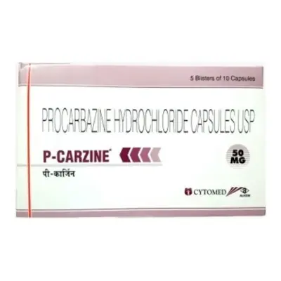
Escalated BEACOPP: An intensified, highly effective regimen (Bleomycin, Etoposide, Doxorubicin, Cyclophosphamide, Vincristine, Procarbazine, and Prednisone) frequently utilized in European centers for high-risk advanced disease, though associated with higher rates of short-term bone marrow suppression and long-term fertility risks.
-
Brentuximab Vedotin + AVD (The ECHELON-1 Regimen): An established, modern frontline option for advanced Hodgkin lymphoma. Brentuximab vedotin is an antibody-drug conjugate that binds directly to the CD30 protein on Reed-Sternberg cells, delivering a potent cellular toxin (MMAE) straight into the cancer cell. Replacing bleomycin with brentuximab vedotin (A+AVD) eliminates the risk of bleomycin-induced pulmonary fibrosis while significantly improving overall survival.
Multimodal Treatment Approaches: Non-Hodgkin Lymphoma
Treatment for Non-Hodgkin lymphoma depends fundamentally on whether the tumor is biologically indolent or aggressive:
1. Aggressive B-Cell Lymphomas (Diffuse Large B-Cell Lymphoma – DLBCL)
DLBCL is an aggressive cancer that requires immediate, full-dose systemic immunochemotherapy with curative intent:
-
R-CHOP (The Global Gold Standard): Combines targeted immunotherapy with multi-agent cytotoxic chemotherapy:
-
Rituximab (R): A chimeric monoclonal antibody that binds directly to the CD20 protein on the surface of B-cells, flagging them for destruction by host natural killer cells and complement pathways.
-
Cyclophosphamide (C): An alkylating agent that cross-links DNA strands.
-
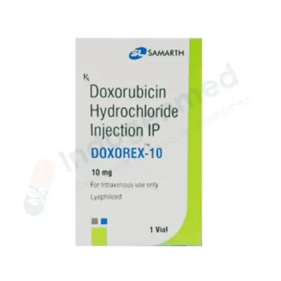
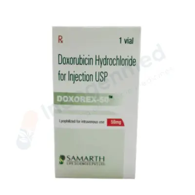
Doxorubicin / Hydroxydaunorubicin (H): An anthracycline that intercalates between DNA base pairs and inhibits topoisomerase II.
-
Vincristine / Oncovin (O): A vinca alkaloid that halts cell division by disrupting the mitotic spindle apparatus.
-
Prednisone (P): An oral corticosteroid that induces apoptosis in lymphoid cells.
-
Administration: R-CHOP is typically administered intravenously every 21 days for 6 cycles. Frontline R-CHOP cures approximately 60% to 70% of all DLBCL patients.
-
Polatuzumab Vedotin + R-CHP (The Pola-R-CHP Regimen): A modern frontline option for intermediate- and high-risk DLBCL. Polatuzumab vedotin is an antibody-drug conjugate targeting CD79b on B-cells. Replacing vincristine with polatuzumab vedotin significantly lowers the risk of disease progression and relapse compared to traditional R-CHOP.
2. Indolent B-Cell Lymphomas (Follicular Lymphoma)
Indolent lymphomas grow slowly, and many patients feel entirely well at diagnosis:
-
Active Surveillance (“Watch-and-Wait”): For asymptomatic patients with low tumor burden, starting chemotherapy immediately does not extend overall survival compared to waiting until symptoms develop. Patients are monitored with physical examinations and blood tests every 3 to 6 months. Active therapy is started only when formal GELF treatment criteria are met (such as the onset of B symptoms, significant organ compression, rapidly enlarging nodes, or dropping blood counts).
-
Frontline Therapies for Active Disease:
-
Obinutuzumab or Rituximab + Bendamustine (BR Regimen): Highly effective, well-tolerated combination delivering high complete response rates with lower hair-loss and nausea rates than older regimens.
-
R-CHOP or R-CVP: Used for bulky or highly symptomatic follicular disease.
-
Maintenance Immunotherapy: Following successful induction chemotherapy, maintenance Rituximab administered once every 2 months for up to 2 years extends progression-free survival.
3. T-Cell and NK-Cell Lymphomas
Peripheral T-cell lymphomas (PTCL) are less common and typically more challenging to treat than B-cell lymphomas:
-
Frontline Combinations: Regimens often incorporate brentuximab vedotin for CD30-positive T-cell lymphomas (such as Anaplastic Large Cell Lymphoma, ALCL) combined with CHP chemotherapy.
-
Consolidation: Due to higher historical recurrence rates, eligible patients with aggressive T-cell lymphomas who achieve complete remission are frequently consolidated with an immediate autologous stem cell transplant to secure long-term remission.
Relapsed and Refractory Disease: Precision Therapies, CAR-T, and Transplants
When lymphoma fails to respond to frontline treatment (refractory disease) or returns after a period of remission (relapsed disease), several second- and third-line therapies are available:
1. High-Dose Chemotherapy and Autologous Stem Cell Transplant (ASCT)
The traditional second-line standard for fit patients with relapsed aggressive DLBCL or Hodgkin lymphoma who demonstrate sensitive disease on second-line salvage chemotherapy:
-
Process: The patient receives salvage chemotherapy to clear bulk tumor. Healthy peripheral blood stem cells are mobilized, collected via an apheresis machine, and frozen. The patient then receives supralethal high-dose conditioning chemotherapy to destroy all remaining microscopic lymphoma cells throughout the body. The thawed stem cells are reinfused intravenously to rescue the bone marrow, allowing blood counts to recover over 2 to 3 weeks in a protective inpatient environment.
2. Chimeric Antigen Receptor (CAR) T-Cell Therapy
One of the most notable therapeutic advancements in modern oncology, CAR T-cell therapy is a personalized cellular immunotherapy approved for relapsed or refractory diffuse large B-cell lymphoma, transformed follicular lymphoma, and mantle cell lymphoma after failing standard therapies:
-
How It Works: A patient’s own T-lymphocytes are harvested from their blood via leukapheresis. In a specialized cell-manufacturing laboratory, a disarmed viral vector introduces a synthetic gene into the T-cells, instructing them to display a Chimeric Antigen Receptor specifically engineered to recognize the CD19 protein on malignant B-cells.
-
Infusion: Following brief conditioning chemotherapy, millions of these living, engineered CAR T-cells are infused back into the patient. Once inside, they multiply exponentially, actively seeking out and destroying CD19-positive lymphoma cells throughout the body.
-
Efficacy: CAR T-cell therapy achieves durable, long-term complete remissions in roughly 35% to 45% of patients with refractory disease who had previously exhausted all conventional chemotherapy options.
3. Bispecific T-Cell Engagers (BiTEs)
Off-the-shelf engineered monoclonal antibodies that feature two separate binding domains:
-
Mechanism: One arm binds specifically to CD20 on malignant lymphoma cells, while the opposite arm binds to CD3 on normal host cytotoxic T-lymphocytes.
-
Action: The bispecific antibody physically pulls the patient’s own T-cells directly into contact with the lymphoma cell, activating the T-cell to inject perforins and granzymes, triggering targeted destruction of the cancer cell. Approved agents include Epcoritamab, Glofitamab, and Mosunetuzumab, providing flexible alternatives for relapsed disease.
4. Oral Targeted Inhibitors
-
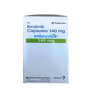
BTK Inhibitors (Ibrutinib, Acalabrutinib, Zanubrutinib): Oral daily tablets that block Bruton’s Tyrosine Kinase, a key enzyme in the B-cell receptor signaling cascade. Highly effective in Mantle Cell Lymphoma, Waldenström’s Macroglobulinemia, and chronic lymphocytic leukemia.
-
PI3K Inhibitors & EZH2 Inhibitors (Tazemetostat): Precision oral tablets that target specific enzymatic mutations in follicular lymphoma.
Survivorship, Long-Term Care, and Managing Treatment Toxicities
As modern treatments continue to cure or manage lymphoma over decades, clinical focus has expanded to address late effects and preserve long-term health:
1. Monitoring for Treatment Toxicities
-
Cardiotoxicity: Anthracyclines (Doxorubicin) can cause cumulative, dose-dependent myocardial strain. Patients undergo baseline echocardiograms to verify Left Ventricular Ejection Fraction (LVEF) before starting therapy, with lifelong monitoring for heart health.
-
Pulmonary Toxicity: Bleomycin can cause oxidative injury to alveolar and interstitial lung tissue, potentially progressing to pulmonary fibrosis. Patients should report any dry cough or shortness of breath; baseline and periodic carbon monoxide diffusing capacity (DLCO) lung tests help identify early toxicity.
-
Peripheral Neuropathy: Vincristine and brentuximab vedotin can cause sensory and motor peripheral neuropathy (numbness, tingling, or weakness in the fingers and toes). Early dose adjustments by the oncology team prevent permanent nerve damage.
-
Secondary Malignancies: Survivors of Hodgkin lymphoma who received chest radiation therapy at a young age face an elevated lifetime risk of secondary breast, lung, or thyroid cancers 10 to 25 years later. Female patients who received thoracic radiation before age 30 should begin annual breast MRI and mammography screening 8 to 10 years after treatment completion (or starting at age 25 to 30).
2. Infection Prevention and Vaccinations
Chemotherapy and anti-CD20 antibodies deplete both normal and malignant B-cells, leading to low antibody levels (hypogammaglobulinemia) that can persist for months after treatment:
-
Prophylaxis: High-risk patients receive daily prophylactic antibiotics (such as trimethoprim-sulfamethoxazole) to prevent Pneumocystis jirovecii pneumonia (PJP), and antiviral medications (acyclovir) to prevent shingles (herpes zoster) reactivation.
-
Vaccines: Patients should receive inactivated seasonal influenza, COVID-19, and pneumococcal vaccines. Live-attenuated vaccines (such as MMR, yellow fever, and oral typhoid) are strictly contraindicated during active immunosuppressive therapy and for at least 6 to 12 months afterward.
3. Fertility Preservation
Because alkylating chemotherapy agents (cyclophosphamide, procarbazine) and pelvic radiation can cause premature ovarian failure or azoospermia:
-
Sperm Cryopreservation: Male patients of reproductive age should be offered sperm banking prior to initiating any systemic therapy.
-
Oocyte or Embryo Cryopreservation: Female patients should receive prompt reproductive endocrinology consultations for emergency egg or embryo harvesting before starting therapy, alongside temporary ovarian suppression using GnRH agonists (goserelin) during chemotherapy cycles to help protect ovarian reserves.
Frequently Asked Questions
Can lymphoma be cured completely?
Yes. Many forms of lymphoma are curable with modern medical care. Classical Hodgkin Lymphoma has long-term cure rates exceeding 85% to 90%. Aggressive Non-Hodgkin lymphomas, such as Diffuse Large B-Cell Lymphoma (DLBCL), achieve complete cures in 60% to 70% of patients with frontline immunochemotherapy (R-CHOP or Pola-R-CHP), and additional patients achieve durable remissions with second-line CAR T-cell therapy. While slow-growing indolent lymphomas (like Follicular Lymphoma) are traditionally considered chronic rather than completely curable, patients often live normal, full lifespans managed with periodic, gentle therapies.
What is the most common first symptom of lymphoma?
The most common initial sign is a painless, firm, swollen lymph node discovered in the neck, above the collarbone, in the armpit, or in the groin. Unlike swollen nodes caused by common infections, lymphoma nodes do not hurt, feel firm or rubbery, and do not shrink back to normal size after 2 to 3 weeks. Other frequent early symptoms include unexplained drenching night sweats, recurring fevers, and unintentional weight loss.
Is an enlarged lymph node always a sign of lymphoma?
No. In fact, the vast majority of swollen lymph nodes are benign and non-cancerous. Lymph nodes act as filtration filters that naturally swell when fighting off common viral or bacterial infections, such as colds, strep throat, ear infections, or dental problems. Benign infection-related nodes are typically soft, tender or painful to the touch, and resolve within a few weeks. A lymph node warrants medical evaluation if it is painless, feels hard or rubbery, measures more than 1 to 2 centimeters in diameter, continues to enlarge, or has been swollen for more than 4 to 6 weeks.
Why is an excisional biopsy better than a needle biopsy for lymphoma?
An excisional biopsy—where a surgeon removes the entire enlarged lymph node intact—is the gold standard because lymphoma diagnosis depends heavily on the internal architectural pattern of the node. A fine-needle aspiration (FNA) draws out only loose individual cells, which cannot show whether cells are growing in follicular circles or diffuse sheets. An intact node provides pathologists with sufficient tissue to perform architectural analysis, extensive immunohistochemical (IHC) staining, flow cytometry, and genetic testing needed to identify the exact lymphoma subtype.
What is the difference between Hodgkin and Non-Hodgkin lymphoma?
The key difference is the presence of a specific abnormal cell. Under a microscope, Hodgkin Lymphoma contains large, abnormal multinucleated cells called Reed-Sternberg cells, which are surrounded by non-cancerous inflammatory immune cells. Hodgkin lymphoma is less common, spreads in an orderly sequence from one node to the next, and is common in young adults. Non-Hodgkin Lymphoma lacks Reed-Sternberg cells, is composed directly of sheets of malignant lymphocytes, is far more common, can develop in any lymphoid or non-lymphoid tissue, and occurs most frequently in adults over age 60.
What are “B Symptoms” and why are they important?
“B Symptoms” refer to three specific systemic warning signs: drenching night sweats that soak clothing, unexplained fevers above 38.0°C (100.4°F), and involuntary loss of more than 10% of body weight over six months. They are caused by high levels of inflammatory cytokines released by the cancer cells or the body’s immune reaction to them. Their presence is recorded as part of the official stage (e.g., Stage IIB) and indicates biologically active disease that typically requires more intensive treatment protocols.
What is CAR T-cell therapy, and who can receive it?
Chimeric Antigen Receptor (CAR) T-cell therapy is a personalized cellular immunotherapy. A patient’s own T-lymphocytes are collected through a blood-filtering process, genetically reprogrammed in a specialized laboratory to produce synthetic surface receptors that target a specific protein on lymphoma cells (such as CD19), and infused back into the patient to hunt and destroy cancer cells. CAR T-cell therapy is primarily approved for patients with aggressive B-cell lymphomas that have relapsed or failed to respond to two or more previous lines of chemotherapy.
What causes lymphoma, and can it be prevented?
In most patients, lymphoma develops from spontaneous, non-inherited genetic errors that occur in lymphocytes during a person’s life, with no single identifiable cause. While there are few modifiable lifestyle risk factors, risk is elevated by chronic immune deficiency (such as HIV or post-transplant immunosuppressants), chronic autoimmune conditions (like Sjögren’s syndrome or celiac disease), certain chronic infections (EBV, Hepatitis C, and H. pylori), and high occupational exposure to industrial solvents or pesticides. Prevention focuses on managing underlying infections, avoiding environmental toxic chemical exposures, and adopting healthy immune-supportive lifestyle habits.
Medical Authority and Clinical Reference Links
-
National Comprehensive Cancer Network (NCCN):
NCCN Clinical Practice Guidelines in Oncology: B-Cell Lymphomas, T-Cell Lymphomas, & Hodgkin Lymphoma.
Consensus clinical practice guidelines for histopathological diagnosis, PET-CT response criteria, targeted therapies, and CAR T-cell therapy:
-
American Society of Hematology (ASH):
ASH Clinical Practice Guidelines and Educational Compendia on Hematologic Malignancies.
Authoritative guidance covering lymphoma diagnosis, immunotherapy toxicities, and stem cell transplantation:
-
European Society for Medical Oncology (ESMO):
ESMO Clinical Practice Guidelines: Diffuse Large B-Cell Lymphoma, Follicular Lymphoma, and Hodgkin Lymphoma.
International recommendations on Lugano staging, molecular biomarker testing, and systemic chemotherapy protocols:
-
World Health Organization (WHO) – International Agency for Research on Cancer (IARC):
WHO Classification of Haematolymphoid Tumours (5th Edition).
The definitive international standard for the pathological classification, genetic definition, and immunohistochemical profiling of lymphoid neoplasms:
-
Leukemia & Lymphoma Society (LLS):
Disease Resources: Hodgkin Lymphoma, Non-Hodgkin Lymphoma, and CAR T-Cell Cellular Therapy.
Biomedical resources detailing subtype biology, clinical trial directories, and patient survivorship protocols: