Stomach Cancer: Symptoms, Causes, Stages, Diagnosis & Treatment
Stomach cancer, clinically termed gastric cancer, is one of the most prevalent and lethal malignancies of the digestive system worldwide. It ranks as the fifth most commonly diagnosed cancer and the fourth leading cause of cancer-related mortality globally, claiming more than 750,000 lives every year.
The disease exhibits marked geographical variation, with exceptionally high incidence rates observed across East Asia, Eastern Europe, and parts of Central and South America.
The overwhelming majority of gastric neoplasms—more than 90% to 95% of all cases—are gastric adenocarcinomas, originating from the mucus-secreting glandular epithelial cells that form the innermost layer of the stomach lining. In Western nations, an anatomical epidemiological shift has occurred over recent decades, characterized by a decrease in distal (non-cardia) gastric tumors and a notable increase in proximal tumors arising at the gastroesophageal junction (GEJ) and the gastric cardia.
Historically associated with poor long-term outcomes due to insidious early presentation, modern clinical management has transformed gastric oncology. Diagnostic precision has improved through high-definition chromoendoscopy, endoscopic ultrasound, and standardized multi-detector staging CT scans. Therapeutically, multimodal perioperative chemotherapy protocols (such as the FLOT regimen), standardized radical gastrectomies with extensive D2 lymph node dissections, and biomarker-driven targeted therapies (anti-HER2, anti-Claudin 18.2, and immune checkpoint inhibitors) have significantly improved curative resections and survival rates.
This comprehensive clinical guide details the anatomy and pathophysiology of the stomach, the Correa cascade of carcinogenesis, clinical warning signs, risk factors, modern diagnostic algorithms, TNM staging, multimodal treatment pathways, and long-term nutritional survivorship.
Key Facts at a Glance
-
Typical Age Demographic: Risk rises sharply after age 50; peak incidence occurs between ages 60 and 80.
-
Gender Predilection: Noticeable male predominance globally, with men affected roughly twice as often as women (2:1 male-to-female ratio).
-
Primary Anatomical Origin: Distal stomach (antrum and pylorus) historically; proximal stomach (cardia and gastroesophageal junction) rising in Western demographics.
-
Primary Histological Type: Gastric Adenocarcinoma (~90%–95%), categorized by the Lauren classification into Intestinal and Diffuse subtypes.
-
Key Actionable Biomarkers: Human Epidermal Growth Factor Receptor 2 (HER2), Programmed Death-Ligand 1 (PD-L1 Combined Positive Score / CPS), Microsatellite Instability (MSI-H / dMMR), and Claudin 18.2 (CLDN18.2).
-
Diagnostic Gold Standard: Upper Gastrointestinal Endoscopy (Esophagogastroduodenoscopy / EGD) with multiple targeted mucosal biopsies.
-
Primary Treatment Modalities: Radical Subtotal or Total Gastrectomy with D2 Lymphadenectomy, Endoscopic Submucosal Dissection (ESD), Perioperative Multi-Agent Chemotherapy (FLOT), Targeted Monoclonal Antibodies (Trastuzumab, Zolbetuximab), and Immunotherapy (Nivolumab, Pembrolizumab).
What Is Stomach Cancer? Anatomy and Histopathology
The stomach is an asymmetrical, muscular digestive organ located in the upper left quadrant of the abdominal cavity directly beneath the diaphragm. It acts as a temporary food reservoir, mechanically churning ingested meals through muscular contractions while initiating chemical protein breakdown via hydrochloric acid and pepsin secretion.
Anatomically, the stomach is divided into five structural regions:
-
The Cardia: The entrance zone immediately distal to the gastroesophageal junction (GEJ), connecting the lower esophagus to the stomach.
-
The Fundus: The dome-shaped, upper superior curvature that expands upward to accommodate gas and swallowed food boluses.
-
The Body (Corpus): The largest, central anatomical segment where the majority of acid-producing parietal cells and pepsinogen-producing chief cells reside.
-
The Antrum: The lower, non-acid-secreting muscular segment responsible for mixing and grinding chyme, containing endocrine G-cells that secrete the hormone gastrin.
-
The Pylorus: The distal, muscular sphincter valve that regulates the metered emptying of processed chyme into the duodenal bulb.
The Histological Layers of the Gastric Wall
The wall of the stomach consists of four concentric tissue coats:
-
Mucosa: The innermost layer consisting of simple columnar glandular epithelium, an underlying lamina propria, and a thin muscularis mucosae layer. Malignant adenocarcinomas arise directly from these glandular cells.
-
Submucosa: Dense irregular connective tissue holding blood vessels, lymphatics, and the submucosal nerve plexus. Tumor penetration through the muscularis mucosae into the submucosa defines an invasive carcinoma capable of lymphatic spread
-
Muscularis Propria: Three robust layers of smooth muscle (inner oblique, middle circular, outer longitudinal) that generate digestive peristalsis.
-
Subserosa and Serosa: The subserosa consists of loose connective tissue, while the serosa (visceral peritoneum) forms the outermost slippery epithelial covering. Tumors that breach the serosa (T4a disease) shed malignant cells directly into the peritoneal cavity, leading to peritoneal carcinomatosis.
The Lauren Histological Classification
Unlike many solid organ malignancies that share uniform histology, gastric adenocarcinomas are categorized into two major subtypes based on the Lauren classification, which differ fundamentally in epidemiology, pathogenesis, and clinical behavior:
-
1. The Intestinal Type:
-
Microscopic Features: Characterized by recognizable glandular structures, tubular architecture, and cohesive malignant epithelial cells lining mucin-containing lumens.
-
Epidemiology: Strongly associated with chronic environmental factors, high-salt diets, and long-standing Helicobacter pylori infection. More prevalent in older male patients and endemic geographic regions.
-
Pathogenesis: Progresses through identifiable, step-wise precancerous stages over decades (the Correa cascade). Carries a comparatively better prognosis.
-
2. The Diffuse Type:
-
Microscopic Features: Characterized by non-cohesive, poorly differentiated cells that lack intercellular adhesions, scattering as single cells or loose clusters throughout the gastric wall without forming glands.
-
Signet Ring Cells: A classic histological hallmark where intracellular mucin pushes the nucleus against the cell membrane, creating a signet-ring appearance under light microscopy.
-
Linitis Plastica (“Leather-Bottle Stomach”): Malignant diffuse cells infiltrate and thicken the entire submucosa and muscularis propria without forming an exophytic intraluminal mass. The stomach wall becomes rigid, thickened, and unable to expand.
-
Epidemiology: Equal sex distribution, frequently seen in younger patients (under age 50), often driven by genetic mutations in the CDH1 (E-cadherin) gene, carrying a significantly more aggressive clinical course and higher rates of early peritoneal dissemination.
Pathogenesis: The Correa Cascade and Molecular Pathways
The development of intestinal-type gastric adenocarcinoma follows a decades-long progression known as the Correa Cascade of Gastric Carcinogenesis:
The Correa Cascade of Gastric Carcinogenesis:
Normal Gastric Mucosa
         │ (Chronic Helicobacter pylori infection & oxidative stress)
         ▼
Chronic Superficial Gastritis
         │ (Persistent inflammatory cytokine release: IL-1β, TNF-α)
         ▼
Chronic Atrophic Gastritis (Loss of parietal & chief cell glandular architecture)
         │ (Achlorhydria & secondary bacterial overgrowth)
         ▼
Intestinal Metaplasia (Replacement of gastric glands with goblet & absorptive enterocytes)
         │ (Accumulation of somatic DNA replication errors)
         ▼
Low-Grade to High-Grade Gastric Epithelial Dysplasia
         │ (Inactivation of TP53, microsatellite instability, or HER2 amplification)
         ▼
Invasive Gastric Adenocarcinoma
1. Helicobacter pylori Infection
Helicobacter pylori is classified as a Group 1 human carcinogen by the World Health Organization, directly responsible for over 75% to 80% of all distal gastric cancers:
-
Colonization: These flagellated, microaerophilic Gram-negative bacteria colonize the gastric mucus layer, utilizing bacterial urease to convert urea into carbon dioxide and ammonia, neutralizing local stomach acid to survive.
-
Bacterial Virulence Factors: Strains expressing the CagA (Cytotoxin-Associated Gene A) and VacA (Vacuolating Cytotoxin A) proteins are significantly more oncogenic. CagA is injected directly into gastric epithelial cells via a bacterial type IV secretion system, where it undergoes phosphorylation, alters cellular polarity, stimulates abnormal proliferation, and degrades intercellular adhesion.
-
Hypochlorhydria: Decades of ongoing active inflammation destroy acid-secreting parietal cells, causing gastric atrophy and hypochlorhydria (loss of stomach acid). The neutral pH allows other bacteria to colonize the stomach, converting dietary nitrates into carcinogenic N-nitroso compounds.
2. The Cancer Genome Atlas (TCGA) Molecular Subtypes
Modern molecular oncology categorizes gastric cancers into four distinct genomic subtypes that direct systemic targeted therapies:
-
Epstein-Barr Virus-Positive (EBV, ~9%): Tumors displaying high frequencies of PIK3CA mutations, extreme DNA hypermethylation (silencing the CDKN2A promoter), and high amplification of CD274 and PDCD1LG2 (which encode PD-L1 and PD-L2). These tumors are sensitive to immune checkpoint inhibitors.
-
Microsatellite Instability-High (MSI-H, ~22%): Tumors characterized by defective DNA mismatch repair proteins (MLH1 hypermethylation), high somatic mutation burdens (high TMB), and prominent lymphocyte infiltration. MSI-H gastric cancers respond to single-agent immunotherapy.
-
Genomically Stable (GS, ~20%): Encompasses predominantly diffuse-type adenocarcinomas and early-onset cancers. Characterized by mutations in the CDH1 (E-cadherin) and RHOA genes, or CLDN18-ARHGAP gene fusions. They lack marked aneuploidy and are resistant to standard chemotherapy.
-
Chromosomal Instability (CIN, ~50%): Encompasses predominantly intestinal-type adenocarcinomas located at the gastroesophageal junction and cardia. Characterized by marked aneuploidy, recurrent TP53 mutations, and focal amplifications of receptor tyrosine kinases—notably ERBB2 (HER2), EGFR, MET, and VEGFR2.
Symptoms and Warning Signs of Stomach Cancer
Early-stage stomach cancer is frequently asymptomatic or causes subtle, non-specific dyspeptic signs that mimic common, benign digestive conditions like gastritis or non-ulcer dyspepsia. Consequently, many patients dismiss symptoms for months before seeking medical evaluation.
1. Early and Subtle Manifestations
-
Persistent Dyspepsia and Indigestion: Mild, nagging upper abdominal discomfort, postprandial fullness, or bloating that fails to resolve with over-the-counter antacids.
-
Epigastric Discomfort: A dull, aching pain localized to the epigastrium that is often unrelated to meals or only partially relieved by eating.
-
Early Satiety: An inability to consume normal-sized meals, feeling full after only a few bites. This occurs when tumors infiltrate and stiffen the gastric wall, reducing the stomach’s natural accommodation capacity.
-
Mild Nausea and Anorexia: A subtle, progressive loss of appetite, often accompanied by a distinct, acquired aversion to protein-rich foods (specifically meat).
2. Progressive and Advanced Symptoms
As the tumor mass enlarges, ulcerates the mucosa, or obstructs the luminal transit of food, clinical signs become more pronounced:
-
Significant Unintentional Weight Loss: Rapid loss of body weight and skeletal muscle wasting (cachexia) driven by reduced caloric intake, malabsorption, and tumor-mediated metabolic inflammation.
-
Chronic Gastrointestinal Bleeding and Anemia: Continual, microscopic bleeding from an ulcerated tumor surface depletes iron reserves, leading to unexplained iron deficiency anemia, profound fatigue, exertional shortness of breath, and pale skin/conjunctiva.
-
Overt Bleeding (Melena and Hematemesis): Passage of black, sticky, tarry stools (melena) caused by digested blood, or the vomiting of bright red blood or dark “coffee-ground” material.
-
Dysphagia (Difficulty Swallowing): Tumors arising in the proximal stomach or cardia encroach upon the gastroesophageal junction, causing food to stick in the lower chest or retrosternal area.
-
Gastric Outlet Obstruction (GOO): Tumors growing in the distal antrum or pylorus mechanically block the exit of the stomach, leading to persistent nausea, upper abdominal distension, and forceful, projectile vomiting of undigested food consumed hours or days earlier.
In advanced, metastatic presentations, physical examination can reveal distinct signs:
-
Virchow’s Node (Troisier’s Sign): A firm, painless, enlarged lymph node palpable in the left supraclavicular fossa, representing retrograde metastatic spread via the thoracic duct.
-
Sister Mary Joseph Nodule: A palpable, hard, violaceous nodule at the umbilicus, indicating transperitoneal tracking along the falciform ligament.
-
Irish’s Node: An isolated, firm, enlarged lymph node in the left axilla.
-
Krukenberg Tumor: Bilateral or unilateral enlargement of the ovaries caused by transcoelomic shedding of signet-ring cell adenocarcinoma cells across the peritoneal cavity into the pelvis.
-
Blumer’s Shelf: A firm, palpable shelf-like mass felt along the anterior rectal wall during a digital rectal examination, reflecting malignant cells pooling and implanting in the rectovesical or rectouterine pouch of Douglas.
-
Malignant Ascites & Hepatomegaly: Abdominal swelling caused by peritoneal metastases, or an irregular, nodular liver edge on palpation caused by extensive hepatic metastases.
Causes and Comprehensive Risk Factor Analysis
Gastric carcinogenesis is a multifactorial process driven by infectious agents, dietary habits, environmental carcinogens, and inherited genetic mutations:
1. Infectious Pathogens
-
Helicobacter pylori: As the primary cause of distal non-cardia gastric cancer, lifelong infection increases the relative risk of developing gastric adenocarcinoma by nearly six-fold. Eradicating H. pylori with targeted antibiotic regimens reduces subsequent cancer incidence, particularly when treated before severe atrophic changes or intestinal metaplasia develop.
-
Epstein-Barr Virus (EBV): The human gammaherpesvirus responsible for mononucleosis is detected in roughly 5% to 10% of all gastric carcinomas worldwide. EBV enters epithelial cells, where its latent viral proteins drive intense promoter hypermethylation that silences host tumor suppressor genes.
2. Dietary and Environmental Influences
-
High-Salt and Salt-Preserved Foods: High consumption of heavily salted, pickled, or cured foods (common in traditional East Asian diets). High concentrations of sodium chloride disrupt the protective gastric mucosal barrier, causing chemical mucosal inflammation, accelerating H. pylori colonization, and increasing gastric epithelial DNA synthesis.
-
Smoked Foods and N-Nitroso Compounds: Regularly consuming smoked foods, charred meats, and cured meats containing nitrates and nitrites. In the hypochlorhydric stomach, nitrates are converted into powerful mutagenic nitrosamines and nitrosamides that induce direct DNA point mutations.
-
Low Consumption of Fresh Produce: Diets lacking fresh fruits, vegetables, and antioxidants (Vitamin C, Vitamin E, carotenoids), which normally scavenge reactive oxygen species and inhibit endogenous nitrosation reactions in gastric fluid.
-
Tobacco Smoking: Active cigarette smoking increases the risk of gastric cancer by 50% to 60%, with carcinogens dissolving into saliva and being repeatedly swallowed, directly contacting the gastric mucosa. Smoking also strongly correlates with adenocarcinomas of the cardia and gastroesophageal junction.
-
Occupational Exposures: Chronic exposure to industrial dusts, including coal mining, asbestos, crystalline silica, and metal refining (nickel and chromium).
3. Inherited Genetic Syndromes
Approximately 1% to 3% of all gastric cancers stem from clearly defined inherited familial cancer syndromes:
-
Hereditary Diffuse Gastric Cancer (HDGC): An autosomal dominant condition caused by germline mutations in the CDH1 gene, which encodes the critical calcium-dependent cellular adhesion glycoprotein E-cadherin.
-
Lifetime Penetrance: Carriers of a pathogenic CDH1 mutation face a 60% to 80% cumulative lifetime risk of developing aggressive, diffuse-type signet-ring cell gastric cancer, along with an elevated risk of lobular breast cancer in women.
-
Prophylactic Gastrectomy: Because microscopic diffuse signet-ring foci develop beneath an intact, normal-looking mucosal surface (making routine endoscopic screening ineffective), clinical consensus guidelines recommend that young adult carriers undergo prophylactic total gastrectomy between ages 20 and 30.
-
Lynch Syndrome (HNPCC): Caused by germline mutations in DNA mismatch repair genes (MLH1, MSH2, MSH6, PMS2), carrying a 5% to 10% lifetime risk of gastric cancer alongside high colorectal and endometrial cancer risks.
-
Familial Adenomatous Polyposis (FAP): Caused by germline mutations in the APC gene; patients develop fundic gland polyps, gastric adenomas, and face an elevated risk of duodenal and gastric carcinomas.
-
Li-Fraumeni Syndrome (TP53) & Peutz-Jeghers Syndrome (STK11): Confer modest elevations in lifetime gastric malignancy risk.
4. Pre-Existing Medical Conditions
-
Autoimmune Metaplastic Atrophic Gastritis (Pernicious Anemia): Autoantibodies destroy gastric parietal cells and intrinsic factor, leading to severe achlorhydria, Vitamin B12 deficiency, hypergastrinemia, and a 3- to 5-fold increased risk of both intestinal-type adenocarcinoma and gastric neuroendocrine (carcinoid) tumors.
-
Prior Partial Gastric Resection (Gastric Stump Carcinoma): Individuals who underwent partial gastrectomy decades earlier for benign peptic ulcer disease face a higher cancer risk in the remaining gastric remnant 15 to 20 years post-surgery, driven by continuous alkaline bile reflux through the surgically altered anastomosis.
-
Menetrier’s Disease: A rare, acquired, hypertrophic gastropathy characterized by massive, giant gastric mucosal folds, severe foveolar hyperplasia, protein-losing enteropathy (hypoalbuminemia), and an elevated risk of malignant transformation.
Diagnostic Evaluation: Endoscopy and Staging Workup
Accurate diagnosis requires direct mucosal visualization, extensive tissue acquisition, and cross-sectional radiological staging:
Diagnostic & Staging Pathway for Gastric Cancer:
[Clinical Suspicion / Red-Flag Dyspeptic Symptoms]
                         │
                         ▼
[Upper Gastrointestinal Endoscopy (EGD) + 6 to 8 Core Biopsies]
                         │
                         ▼
[Histopathology Confirmation: Lauren Classification & Biomarkers]
(HER2 IHC/FISH, PD-L1 CPS, MSI/MMR, Claudin 18.2)
                         │
                         ▼
[Multi-Detector Dynamic CT of Chest, Abdomen, and Pelvis]
                         │
         ┌───────────────┴───────────────┐
         ▼                               ▼
[Metastatic Disease Confirmed]   [Localized / Locally Advanced Disease]
         │                               │
         ▼                               ▼
[Palliative Systemic Therapy]    [Endoscopic Ultrasound (EUS)]
                                 (Assess T-Depth & N-Nodal Staging)
                                                 │
                                                 ▼
                                 [Diagnostic Staging Laparoscopy + Peritoneal Washings]
                                 (Rule out occult sub-diaphragmatic or peritoneal spread)
1. Upper Gastrointestinal Endoscopy (EGD)
The primary diagnostic tool. A flexible video endoscope is advanced through the esophagus, stomach, and duodenal loop:
-
Visual Assessment: Gastric cancers present as flat mucosal discolorations, shallow depressed lesions, malignant non-healing ulcers with heaped-up, irregular margins, or large exophytic fungating masses.
-
Biopsy Protocol: International guidelines mandate obtaining at least 6 to 8 separate tissue biopsies from the ulcer margins and center. A single biopsy carries a 70% diagnostic sensitivity, whereas taking 6 to 8 biopsies raises histological diagnostic accuracy to over 98%.
-
Image-Enhanced Endoscopy: High-definition endoscopes equipped with Narrow Band Imaging (NBI) or chromoendoscopy (spraying dyes like indigo carmine or acetic acid onto the mucosa) highlight abnormal microvascular networks and micro-surface mucosal patterns, guiding targeted biopsies of subtle, early lesions.
2. Mandatory Biomarker Profiling
All biopsy specimens of gastric and gastroesophageal junction adenocarcinomas must undergo formal biomarker testing to guide precision systemic therapies:
-
HER2 Status (ERBB2): Tested via Immunohistochemistry (IHC) with reflex Fluorescence In Situ Hybridization (FISH) for equivocal cases. Positive overexpression with FISH amplification) occurs in roughly 15% to 20% of gastric tumors (predominantly intestinal-type at the GEJ). HER2 positivity directs the addition of Trastuzumab to frontline chemotherapy.
-
PD-L1 Expression (Combined Positive Score – CPS): Measures the number of PD-L1-staining cells (tumor cells, lymphocytes, macrophages) relative to the total number of viable tumor cells:
-
Mismatch Repair (MMR) / Microsatellite Instability (MSI): Evaluated via IHC for the four mismatch repair proteins (MLH1, MSH2, MSH6, PMS2). dMMR/MSI-H tumors demonstrate sensitivity to immunotherapy.
-
Claudin 18.2 (CLDN18.2): A tight-junction protein expressed in gastric mucosal cells that remains exposed on the cell surface during malignant transformation. Strong, moderate-to-high membranous expression in 75% of tumor cells directs the use of Zolbetuximab, a targeted monoclonal antibody.
3. Cross-Sectional Staging Imaging
-
Multi-Detector Contrast-Enhanced CT (Chest, Abdomen, and Pelvis): Thin-slice helical CT performed with both intravenous iodinated contrast and oral water contrast (acting as a negative oral contrast agent to distend the stomach wall). It evaluates primary tumor thickness, local invasion into adjacent organs (pancreas, liver, colon), regional lymph node basins, and screens for distant liver or pulmonary metastases.
-
Endoscopic Ultrasound (EUS): The most accurate modality for determining local anatomical depth of invasion (T-stage) and regional lymph node involvement (N-stage). A high-frequency ultrasound transducer visualizes the distinct five-layer structure of the gastric wall, reliably distinguishing superficial mucosal lesions (amenable to endoscopic resection) from deep muscularis or serosal invasion.
4. Diagnostic Staging Laparoscopy with Peritoneal Cytology
Mandatory for all patients with locally advanced gastric cancer (cT3 or cT4, or any node-positive disease) being considered for curative-intent surgery.
-
The Clinical Rationale: Multi-detector CT scans miss up to 20% to 30% of small, microscopic peritoneal metastases (sub-centimeter “miliary” nodules scattered across the omentum, mesentery, and diaphragm).
-
The Procedure: A surgeon inserts a laparoscope into the peritoneal cavity to directly inspect the liver surfaces, omentum, and pelvic peritoneal reflection.
-
Peritoneal Washings: Saline is instilled into the subphrenic space and pelvis, aspirated, and sent for cytological examination. The presence of free malignant cells in peritoneal washings (even without visible macroscopic tumors) is classified as metastatic disease (M1), making radical surgery ineffective and directing treatment toward systemic chemotherapy.
Staging of Stomach Cancer: The AJCC TNM System
Stomach cancer is staged using the American Joint Committee on Cancer (AJCC) TNM 8th Edition framework:
Clinical Stage Groupings and Prognostic Profile
-
Stage I (T1N0-1, T2N0): Early, localized disease; five-year survival exceeds 70% to 90% with surgical resection.
-
Stage II (T1N2-3, T2N1-2, T3N0-1, T4aN0): Moderately advanced local disease with early nodal involvement; managed via perioperative chemotherapy combined with radical surgery.
-
Stage III (T3-4 with heavy N-nodal spread): Locally advanced, deep invasion with extensive regional lymph node metastasis, requiring intensive multimodal care.
-
Stage IV (Any T, Any N, M1): Distant metastatic dissemination; managed primarily through systemic chemotherapy, targeted biologics, and supportive palliative care.
Multimodal Treatment Approaches: Early and Localized Disease
Treating localized gastric cancer requires an integrated multimodal plan determined by a multidisciplinary team (surgical oncologists, medical oncologists, gastroenterologists, and pathologists):
Multimodal Treatment Strategy for Gastric Cancer:
[Very Early Lesions (cT1a, Well-Differentiated, < 2 cm, No Ulcer)]
                         │
                         ▼
[Endoscopic Submucosal Dissection (ESD / EMR) - Organ Preserved]

[Locally Advanced Resectable Disease (≥ cT2 or Node-Positive)]
                         │
                         ▼
[Neoadjuvant Chemotherapy: FLOT Regimen (4 Cycles Prior to Surgery)]
                         │
                         ▼
[Radical Gastrectomy + Standard D2 Lymphadenectomy (Subtotal vs. Total)]
                         │
                         ▼
[Adjuvant Chemotherapy: FLOT Regimen (4 Cycles Post-Surgery)]
1. Endoscopic Resection for Early Gastric Cancer (EGC)
For very early cancers confined strictly to the superficial mucosa:
-
Endoscopic Submucosal Dissection (ESD): A minimally invasive, organ-sparing endoscopic procedure. Under conscious sedation, an endoscopist injects fluid into the submucosal space to lift the lesion away from the muscularis propria, then uses a micro-electrosurgical knife to dissect the entire lesion free in one intact piece (en bloc).
-
Standard Indications: Differentiated adenocarcinoma, tumor size, strictly confined to the mucosa (T1a), lacking ulceration, and with no evidence of lymphovascular space invasion.
-
Oncological Outcome: Provides a 5-year survival rate exceeding 95% with zero risk of post-gastrectomy digestive complications, while avoiding major abdominal surgery.
2. Perioperative Systemic Chemotherapy (The FLOT Protocol)
For medically fit patients presenting with localized, resectable gastric cancer that is Stage T2 or higher, or has any regional lymph node involvement:
-
The FLOT Regimen: The established international standard of care, supported by the landmark German AIO-FLOT4 trial. A triplet combination consisting of:
-
5-Fluorouracil (5-FU): Infused continuously over 24 hours.
-
Leucovorin: Biochemical modulator enhancing 5-FU binding.
-
Oxaliplatin: Platinum DNA cross-linking agent.
-
Docetaxel: Microtubule-stabilizing taxane chemotherapy.
-
The Perioperative Schedule: Delivered as 4 cycles before surgery (neoadjuvant) and 4 cycles after surgery (adjuvant), administered every 2 weeks.
-
Clinical Benefits: Neoadjuvant FLOT treats microscopic metastatic disease early, achieves tumor downstaging, significantly increases the rate of complete microscopically negative surgical resections, and delivers pathological complete response (pCR) rates of roughly 15% to 20%, translating into superior overall survival compared to surgery followed by postoperative chemotherapy alone.
Surgical Oncology: Gastrectomy and D2 Lymphadenectomy
Complete surgical removal of the tumor with clear margins and radical lymph node dissection is the cornerstone of curative therapy for operable gastric cancer:
1. Types of Gastric Resections
The location of the primary tumor dictates the type of gastrectomy performed:
-
Subtotal (Distal) Gastrectomy: Indicated for tumors located in the lower stomach (antrum or pylorus). The surgeon resects the distal two-thirds of the stomach along with the pylorus, omentum, and regional lymph nodes, preserving the upper stomach fundus and cardia. Preserving the proximal gastric reservoir supports post-operative nutrition and reduces dumping syndrome. Digestive continuity is restored via a Roux-en-Y gastrojejunostomy or Billroth II reconstruction.
-
Total Gastrectomy: Mandatory for tumors located in the upper or middle stomach (body, fundus, or cardia), or for diffuse-type linitis plastica involving multiple gastric regions. The entire stomach is resected from the distal esophagus to the duodenal bulb, along with the greater and lesser omenta.
-
Reconstruction: Reconstructed via a Roux-en-Y Esophagojejunostomy, where a loop of mid-jejunum is mobilized and sewn directly to the distal esophagus, forming a direct conduit for food transit to the small intestine.
-
Clear Margins: In intestinal-type cancers, a minimum microscopic proximal margin of 3 to 5 centimeters is required. In diffuse-type cancers, because malignant cells infiltrate invisibly along submucosal planes, wide gross margins of at least 5 to 8 centimeters are necessary to prevent positive surgical margins.
2. The Extent of Lymphadenectomy: D1 vs. D2 Dissection
Gastric cancer metastasizes early to regional lymph node basins arranged in concentric stations surrounding the stomach:
-
D1 Lymphadenectomy: Removes only the perigastric lymph nodes located immediately adjacent to the gastric walls (Stations 1 through 6).
-
D2 Lymphadenectomy: Removes all D1 perigastric nodes plus the second-tier lymph nodes traveling along the main branches of the celiac axis: the left gastric artery (Station 7), common hepatic artery (Station 8), celiac trunk (Station 9), splenic artery (Station 11), and the splenic hilum (Station 10 in proximal tumors).
-
The Standard of Care: Long-term follow-up from the Dutch Gastric Cancer Trial confirmed that D2 lymphadenectomy is the global surgical standard of care for curative-intent resections. When performed by experienced surgeons in high-volume centers, D2 dissection significantly reduces local and regional recurrence rates and improves overall cancer survival without increasing operative mortality. International guidelines mandate that at least 16, and ideally 30 or more, lymph nodes must be resected and histopathologically examined for accurate nodal staging.
For patients presenting with unresectable, locally advanced, or distant metastatic gastric cancer (Stage IV), treatment centers on biomarker-directed systemic therapies designed to control disease, relieve symptoms, and prolong survival:
Biomarker-Driven First-Line Metastatic Strategy:
                                Advanced / Metastatic Gastric Cancer
                                                  │
                            ┌─────────────────────┴─────────────────────┐
                            ▼                                           ▼
                    [HER2-Positive]                             [HER2-Negative]
                            │                                           │
                            ▼                                           ▼
             [Platinum + Fluoropyrimidine]               [Evaluate PD-L1 CPS & Claudin 18.2]
                            +                                           │
                      [Trastuzumab]                      ┌──────────────┴──────────────┐
                            +                            ▼                             ▼
                    [Pembrolizumab]               [PD-L1 CPS ≥ 5]             [Claudin 18.2 Positive]
               (If PD-L1 CPS is positive)                │                             │
                                                         ▼                             ▼
                                           [Platinum + Fluoropyrimidine] [mFOLFOX / CAPOX]
                                                         +                             +
                                                    [Nivolumab]                 [Zolbetuximab]
1. Frontline HER2-Positive Gastric Cancer
For patients whose tumors overexpress the HER2 protein:
-
The Standard Regimen: A chemotherapy doublet of a fluoropyrimidine (Capecitabine or 5-FU) and a platinum agent (Oxaliplatin or Cisplatin) combined with Trastuzumab, an anti-HER2 monoclonal antibody.
-
Adding Immunotherapy: Results from the phase III KEYNOTE-811 trial demonstrated that adding the immune checkpoint inhibitor Pembrolizumab to frontline Trastuzumab and chemotherapy significantly increases objective response rates and extends progression-free survival in patients whose tumors are both HER2-positive and PD-L1-positive.
2. Frontline HER2-Negative Gastric Cancer
For the majority of patients with HER2-negative disease:
-
PD-L1 Expression Driven (CheckMate-649 Protocol): In patients whose tumors demonstrate a PD-L1 Combined Positive Score (CPS), the international standard of care combines systemic chemotherapy (FOLFOX or CAPOX) with Nivolumab, an anti-PD-1 immune checkpoint inhibitor. This combination significantly extends overall survival compared to chemotherapy alone.
-
Claudin 18.2 Targeted Therapy (SPOTLIGHT and GLOW Trials): For patients with HER2-negative, Claudin 18.2-positive tumors (defined as moderate-to-strong membranous expression in 75% of tumor cells), adding Zolbetuximab (a chimeric monoclonal antibody that binds Claudin 18.2 and induces antibody-dependent cellular cytotoxicity) to frontline mFOLFOX6 or CAPOX significantly improves progression-free and overall survival.
3. Second-Line Targeted Regimens
When cancer progresses following first-line therapy:
-
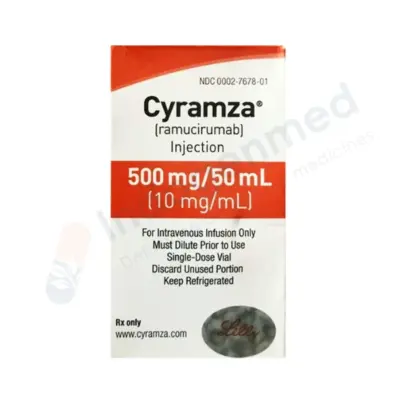
Ramucirumab + Paclitaxel (The RAINBOW Protocol): Ramucirumab is a fully human monoclonal antibody that binds to Vascular Endothelial Growth Factor Receptor-2 (VEGFR-2), blocking tumor angiogenesis. Combined with weekly paclitaxel, it represents the primary international standard for second-line therapy.
-
Trastuzumab Deruxtecan (T-DXd): An advanced antibody-drug conjugate approved for patients with HER2-positive gastric cancer who have progressed on initial trastuzumab-based regimens. It delivers a topoisomerase I inhibitor payload directly into HER2-expressing cells, overcoming acquired resistance.
Post-Gastrectomy Care and Long-Term Nutritional Survivorship
Resecting part or all of the stomach fundamentally alters normal gastrointestinal anatomy and digestive physiology. Long-term survivorship requires proactive nutritional management:
1. Managing Post-Gastrectomy Syndromes
-
Dumping Syndrome: Caused by the rapid transit of concentrated, hyperosmolar food into the proximal small intestine:
-
Early Dumping (within 15 to 30 minutes post-meal): Rapid fluid shifts from the bloodstream into the bowel lumen cause sudden abdominal cramping, nausea, explosive diarrhea, dizziness, facial flushing, and tachycardia.
-
Late Dumping (1 to 3 hours post-meal): Rapid carbohydrate absorption triggers an exaggerated insulin spike, resulting in reactive hypoglycemia (sweating, tremors, confusion, hunger, weakness).
-
Dietary Management: Consume small, frequent meals (6 to 8 meals daily); avoid refined sugars and simple sweets; consume liquids 30 to 45 minutes after solid meals rather than during eating; increase dietary soluble fiber and complex carbohydrates.
-
Alkaline Reflux Gastritis / Esophagitis: Retrograde reflux of bile and pancreatic secretions from the jejunum into the lower esophagus, causing burning chest pain and bilious vomiting; managed via Roux-en-Y surgical reconstruction, which diverts bile drainage 40 to 50 cm downstream from the esophageal anastomosis.
2. Essential Micronutrient and Vitamin Replacements
Following total gastrectomy, lifelong oral and injectable supplementation is mandatory:
-
Vitamin B12 (Cobalamin): Total gastrectomy completely removes all gastric parietal cells, eliminating intrinsic factor production. Without intrinsic factor, physiological absorption of dietary Vitamin B12 in the terminal ileum drops to zero. All total gastrectomy patients require lifelong intramuscular Vitamin B12 injections (1,000 mcg administered every 1 to 3 months) to prevent megaloblastic anemia and subacute combined spinal cord degeneration.
-
Iron Deficiency: Normal iron absorption requires gastric acid to reduce dietary ferric iron to the soluble ferrous state for duodenal uptake. Bypassing the duodenum and stomach leads to chronic iron deficiency, requiring daily oral iron formulations or periodic intravenous iron infusions.
-
Calcium and Vitamin D: Calcium absorption is reduced by duodenal bypass and rapid intestinal transit, accelerating bone mineral loss and osteopenia. Patients require regular bone density monitoring and daily supplemental calcium citrate paired with Vitamin D3.
-
Weight Stabilization and Pancreatic Enzymes: Patients frequently experience post-gastrectomy malabsorption and weight loss. Oral Pancreatic Enzyme Replacement Therapy (PERT) can be prescribed to support fat digestion and improve nutrient absorption.
Prevention, Screening, and Eradication Strategies
-
Helicobacter pylori Eradication: Population-level testing and treatment of H. pylori is the most effective intervention for reducing distal gastric cancer incidence. Quadruple therapy regimens—combining a high-dose Proton Pump Inhibitor (or Potassium-Competitive Acid Blocker), Bismuth Subsalicylate, Metronidazole, and Tetracycline for 14 days—achieve high eradication rates, eliminating chronic mucosal inflammation.
-
Endoscopic Screening in High-Incidence Regions: In endemic countries (South Korea, Japan), universal national screening programs utilizing biennial screening upper endoscopies for all adults aged 40 and older detect over 60% to 70% of gastric cancers at early Stage I (EGC), allowing curative endoscopic resection and dramatically lowering population mortality.
-
Dietary Adjustments: Lowering consumption of salt-cured, pickled, and chemically smoked foods, eliminating processed meats containing sodium nitrite, avoiding tobacco products, and increasing daily intake of fresh fruits, leafy vegetables, and dietary citrus fruits rich in Vitamin C.
-
Targeted Surveillance for Premalignant Conditions: Patients with confirmed high-risk conditions—such as extensive gastric intestinal metaplasia, severe atrophic gastritis, or familial polyposis syndromes—should undergo structured surveillance endoscopies with high-definition chromoendoscopy every 1 to 3 years.
Which Medical Specialist Should You Consult?
A comprehensive medical evaluation coordinates accurate diagnostic workup, endoscopic staging, and treatment planning:
-
Gastroenterologist: The primary specialist who performs upper endoscopies (EGD), visualizes suspicious mucosal lesions, takes diagnostic biopsies, and executes Endoscopic Submucosal Dissection (ESD) for early-stage mucosal cancers.
-
Surgical Oncologist / Hepato-Pancreato-Biliary & Upper GI Surgeon: A fellowship-trained surgeon specializing in oncological resections of the upper digestive tract, performing subtotal gastrectomies, total gastrectomies with radical D2 lymphadenectomy, and diagnostic staging laparoscopies.
-
Medical Oncologist: Specializes in systemic therapies, managing perioperative chemotherapy (the FLOT protocol), targeted monoclonal antibodies (Trastuzumab, Zolbetuximab), anti-angiogenic agents, and checkpoint immunotherapy.
-
Gastrointestinal Pathologist: Analyzes biopsy and surgical resection specimens, determining histological subtype (Lauren classification), checking surgical margins, counting resected lymph nodes, and conducting biomarker testing (HER2, PD-L1 CPS, MSI/MMR, Claudin 18.2).
-
Oncology Clinical Dietitian: Essential for managing post-gastrectomy nutrition, designing low-glycemic meal plans to prevent dumping syndrome, monitoring micronutrient levels, and coordinating oral pancreatic enzyme replacements.
Frequently Asked Questions
Can stomach cancer be cured completely?
Yes. Stomach cancer can be cured completely if it is diagnosed and treated at an early, localized stage. Early gastric cancers confined to the superficial mucosal layer can be cured with minimally invasive Endoscopic Submucosal Dissection (ESD), delivering five-year survival rates exceeding 95%. For locally advanced cancers that have invaded into the muscular wall or nearby lymph nodes, combining perioperative chemotherapy (the FLOT regimen) with a radical subtotal or total gastrectomy and D2 lymph node dissection offers a realistic chance of long-term cure. However, cure is rare once cancer has spread to distant organs or seeded the peritoneal cavity.
What is the most common early symptom of stomach cancer?
Early stomach cancer rarely causes dramatic symptoms; its earliest signs are subtle and easily mistaken for common indigestion, mild gastritis, or acid reflux. The most frequent early complaints include persistent upper abdominal discomfort, a sensation of fullness or bloating after eating small amounts of food (early satiety), mild nausea, and a gradual loss of appetite. Any persistent indigestion or upper abdominal pain lasting more than two to three weeks in an adult over age 45 to 50 warrants medical evaluation with an upper endoscopy.
How does Helicobacter pylori bacteria cause stomach cancer?
Helicobacter pylori is a spiral-shaped bacterium that lives in the acidic stomach environment by using the enzyme urease to neutralize acid around itself. It adheres to gastric cells and injects inflammatory toxins (such as CagA), causing chronic, lifelong inflammation of the stomach lining (chronic gastritis). Over decades, this continuous inflammation destroys acid-producing glands (atrophic gastritis), replaces normal stomach cells with intestinal-type cells (intestinal metaplasia), and promotes DNA mutations that can progress into invasive cancer.
What is the difference between a partial (subtotal) and a total gastrectomy?
A partial (subtotal) gastrectomy removes the lower two-thirds of the stomach, leaving the upper portion intact; it is performed for tumors located in the lower stomach (antrum or pylorus). A total gastrectomy involves the surgical removal of the entire stomach from the lower esophagus to the beginning of the small intestine; it is required for tumors located in the middle or upper stomach, or for diffuse-type cancers that spread throughout the gastric wall. Following total gastrectomy, the surgeon connects the esophagus directly to the small intestine (jejunum).
Can a person live a normal life without a stomach?
Yes, a person can live a full, functional life without a stomach. Because the stomach’s main roles are holding food and initiating digestion, the small intestine adapts over time to absorb nutrients directly. Patients who have had a total gastrectomy must adapt their eating habits by consuming 6 to 8 small, nutrient-dense meals throughout the day rather than two or three large meals, chewing food thoroughly, and drinking fluids between meals rather than with food. They also require lifelong intramuscular Vitamin B12 injections and regular iron and calcium supplements.
What is “Dumping Syndrome” and how is it managed?
Dumping syndrome is a group of digestive symptoms that occur when food—especially sugars and refined carbohydrates—empties too rapidly from the esophagus or stomach remnant directly into the small intestine. Early dumping occurs within 30 minutes of eating, causing cramping, nausea, diarrhea, facial flushing, and a racing heart. Late dumping occurs 1 to 3 hours later, causing shakiness, sweating, and weakness resulting from reactive low blood sugar (hypoglycemia). It is managed by eating small, frequent, low-carbohydrate meals, increasing dietary soluble fiber, and avoiding drinking liquids during meals.
What is the difference between intestinal and diffuse stomach cancer?
Under the Lauren classification, intestinal-type stomach cancer forms organized, recognizable glandular structures; it is more common in older men, linked to environmental factors like diet and H. pylori, and follows a slow, step-wise progression. Diffuse-type stomach cancer consists of scattered, individual cancer cells (including signet-ring cells) that infiltrate through the layers of the stomach wall without forming a discrete mass, making the stomach rigid (linitis plastica). Diffuse stomach cancer is seen equally in men and women, often affects younger individuals, is frequently linked to genetic CDH1 mutations, and carries a more aggressive clinical course.
What is HER2 testing in stomach cancer, and why is it important?
HER2 is a protein receptor found on the surface of some cancer cells that signals the cell to grow and divide rapidly. Approximately 15% to 20% of gastric adenocarcinomas produce excessive amounts of HER2 (HER2-positive). Every patient diagnosed with advanced or metastatic stomach cancer should have their biopsy tissue tested for HER2. If positive, adding the targeted antibody drug Trastuzumab to standard chemotherapy significantly improves tumor shrinkage and extends survival.
Medical Authority and Clinical Reference Links
-
National Comprehensive Cancer Network (NCCN):
NCCN Clinical Practice Guidelines in Oncology: Gastric Cancer.
Consensus clinical practice guidelines covering endoscopic staging, D2 surgical standards, perioperative chemotherapy, and targeted therapies:
-
European Society for Medical Oncology (ESMO):
Gastric Cancer: ESMO Clinical Practice Guidelines for Diagnosis, Treatment, and Follow-up.
International recommendations on molecular classification, perioperative FLOT administration, and biomarker testing:
-
American Society of Clinical Oncology (ASCO):
Treatment of Locally Advanced and Metastatic Gastric Cancer: ASCO Guideline.
Evidence-based consensus guidelines on systemic chemotherapy combinations, anti-HER2 therapies, and immune checkpoint inhibition:
-
World Health Organization (WHO) – International Agency for Research on Cancer (IARC):
H. pylori Eradication as a Strategy for Preventing Gastric Cancer & Global Cancer Statistics (GLOBOCAN):
-
International Gastric Cancer Association (IGCA) & Japanese Gastric Cancer Association (JGCA):
Japanese Gastric Cancer Treatment Guidelines: Staging Classifications and Lymph Node Station Mappings: