Pancreatic Cancer: Symptoms, Causes, Stages, Diagnosis & Treatment
Pancreatic cancer is one of the most aggressive and biologically challenging malignancies in human oncology. Ranking as the third leading cause of cancer-related mortality in many parts of the world despite accounting for a smaller percentage of overall cancer diagnoses, it is characterized by rapid local invasion, an early propensity for distant micrometastasis, and a dense, fibrotic tumor environment that acts as a natural barrier to drug delivery.
The pancreas sits deep within the retroperitoneum, nestled behind the stomach and surrounded by critical vascular structures, including the superior mesenteric artery, superior mesenteric vein, portal vein, and celiac axis. Because the pancreas lacks a true serosal capsule and is located near these major vessels, tumors often invade surrounding tissues early. Furthermore, early-stage pancreatic neoplasms produce few, if any, noticeable symptoms, meaning that more than 80% of patients present with locally advanced or distant metastatic disease at the time of diagnosis.
Over the past decade, advancements in multi-detector cross-sectional imaging, endoscopic ultrasound-guided tissue acquisition, neoadjuvant systemic chemotherapy regimens, and complex arterial/venous vascular resections have shifted clinical paradigms. Patients once considered untreatable can increasingly achieve disease downstaging, potential surgical resections, and prolonged survival.
This comprehensive clinical guide reviews pancreatic anatomy, exocrine versus endocrine tumor biology, clinical warning signs, risk factors, modern diagnostic algorithms, resectability criteria, multimodal treatment protocols, and supportive symptom care.
Key Facts at a Glance
-
Typical Age Demographic: Risk rises progressively after age 50; peak incidence occurs between ages 65 and 80.
-
Gender Predilection: Slightly higher incidence in men compared to women (roughly 1.3 to 1 ratio).
-
Primary Anatomical Origin: The head and uncinate process of the pancreas (accounting for roughly 65% to 70% of primary tumors); the body and tail account for 20% to 25%.
-
Primary Histological Subtype: Pancreatic Ductal Adenocarcinoma (PDAC), originating from the epithelial lining of the exocrine pancreatic ducts, accounting for over 90% of cases.
-
Key Tumor Biomarkers: Serum Carbohydrate Antigen 19-9 (CA 19-9), Carcinoembryonic Antigen (CEA).
-
Primary Diagnostic Modalities: Multi-Phase Contrast-Enhanced Pancreatic-Protocol CT, Contrast MRI/MRCP, Endoscopic Ultrasound with Fine-Needle Biopsy (EUS-FNB).
-
Core Treatment Modalities: Pancreaticoduodenectomy (Whipple procedure), Distal Pancreatectomy, Multi-Agent Chemotherapy (mFOLFIRINOX, Gemcitabine + Nab-Paclitaxel), Stereotactic Body Radiation Therapy (SBRT), PARP inhibitors for BRCA carriers.
What Is Pancreatic Cancer? Anatomy and Pathophysiology
The pancreas is an elongated, retroperitoneal organ measuring approximately 15 centimeters in length, situated horizontally across the posterior abdominal wall. Anatomically, it is divided into five regions: the head, uncinate process, neck, body, and tail. The head rests within the C-shaped curve of the duodenum, while the tail extends laterally to touch the hilum of the spleen.
The pancreas functions as a dual-action organ, housing two distinct cellular compartments:
-
The Exocrine Pancreas (Over 95% of Total Tissue Mass):
-
Composed of acinar cells and a branching ductal network.
-
Function: Synthesizes, stores, and secretes roughly 1.5 to 2 liters of alkaline digestive fluid daily. This fluid contains bicarbonate and digestive enzymes (lipase, amylase, trypsin, chymotrypsin) that travel down the main pancreatic duct (duct of Wirsung) and empty through the ampulla of Vater into the duodenum to digest fats, carbohydrates, and proteins.
-
Pathology: Malignancies arising in this compartment are exocrine tumors, dominated by Pancreatic Ductal Adenocarcinoma (PDAC).
-
The Endocrine Pancreas (Less than 2% to 5% of Tissue Mass):
-
Composed of the Islets of Langerhans scattered throughout the parenchymal tissue, predominantly in the body and tail.
-
Function: Releases systemic regulatory hormones directly into the bloodstream, including insulin (beta cells), glucagon (alpha cells), and somatostatin (delta cells) to manage glucose homeostasis.
-
Pathology: Malignancies arising here are Pancreatic Neuroendocrine Tumors (PanNETs or Islet Cell Tumors). PanNETs behave differently from adenocarcinomas, displaying slower growth rates, distinct genetic drivers, and unique systemic therapies.
The Biology of Pancreatic Ductal Adenocarcinoma (PDAC)
PDAC develops through a stepwise progression of non-invasive microscopic precursors termed Pancreatic Intraepithelial Neoplasia (PanIN), grading from PanIN-1 to high-grade PanIN-3 (carcinoma in situ) before breaching the basement membrane.
A defining characteristic of PDAC is its prominent desmoplastic stroma. The tumor mass consists of a dense, fibrous extracellular matrix populated by cancer-associated fibroblasts (CAFs), myofibroblasts, and collagen fibers. This dense stroma compresses blood vessels, creating high internal fluid pressures and a hypoxic environment. This altered tissue architecture restricts the delivery of intravenous chemotherapy drugs and limits the infiltration of host cytotoxic T-lymphocytes, contributing to the tumor’s resistance to many standard treatments.
At the molecular level, PDAC is driven by four key genetic alterations:
-
KRAS Activation: Point mutations in the KRAS oncogene (most commonly at codon 12) occur in more than 90% to 95% of all PDAC cases, locking the enzyme in an active state that drives cellular division.
-
CDKN2A / p16 Inactivation: Lost in over 90% of tumors, disabling normal cell-cycle arrest.
-
TP53 Inactivation: Mutated or lost in 70% to 80% of tumors, preventing programmed cell death (apoptosis) in response to DNA damage.
-
SMAD4 / DPC4 Inactivation: Inactivated in approximately 50% of tumors, associated with higher rates of widespread metastatic dissemination.
Symptoms and Warning Signs of Pancreatic Cancer
Pancreatic cancer is notoriously insidious. In its early, resectable stages, tumors produce subtle, non-specific gastrointestinal symptoms that are easily attributed to benign conditions such as acid reflux, indigestion, irritable bowel syndrome, or muscular back strain.
Symptoms vary according to where the tumor is located:
1. Symptoms of Tumors in the Pancreatic Head (65% to 70% of Cases)
Because the head of the pancreas wraps around the distal common bile duct as it travels toward the duodenum, tumors here compress the bile duct early in their course:
-
Painless Obstructive Jaundice: The classic warning sign. A yellowing of the skin and eyes (scleral icterus) occurring without significant abdominal pain.
-
Pruritus (Intense Skin Itching): Bile salts cannot drain into the intestine and back up into the bloodstream, depositing in the skin.
-
Dark “Tea-Colored” Urine: Water-soluble conjugated bilirubin spills into the urine.
-
Acholuric (Pale / Clay-Colored) Stools: The stool loses its normal brown color (derived from stercobilin) because bile cannot reach the digestive tract.
-
Courvoisier’s Sign: A palpable, enlarged, non-tender gallbladder discovered in a jaundiced patient, indicating chronic distal bile duct obstruction from a tumor rather than gallstone impaction.
2. Symptoms of Tumors in the Body and Tail (20% to 25% of Cases)
Tumors in the body and tail do not compress the common bile duct. Consequently, they grow to substantial sizes, invade local nerves, or metastasize before producing symptoms:
-
Persistent Epigastric and Mid-Back Pain: The classic pain of pancreatic cancer is a dull, deep, unremitting ache in the upper abdomen that radiates straight through to the mid-back. The pain classically worsens after eating and when lying flat on one’s back (supine), and eases when leaning forward or curling into a fetal position, which relieves pressure off the retroperitoneal celiac plexus nerves.
-
Severe Unexplained Weight Loss and Anorexia: Rapid loss of body weight and profound skeletal muscle wasting (cancer cachexia).
-
Exocrine Pancreatic Insufficiency (EPI): The tumor blocks pancreatic enzymes from reaching the duodenum, preventing normal fat breakdown. This produces steatorrhea—large, foul-smelling, pale, greasy stools that float and are difficult to flush—accompanied by bloating, cramping, and fat-soluble vitamin deficiencies (Vitamins A, D, E, and K).
-
New-Onset Type 2 Diabetes: The sudden onset of type 2 diabetes in an adult over age 50 who lacks typical risk factors (such as a family history of diabetes or obesity), or a sudden, unexplained deterioration in blood sugar control in a previously stable diabetic patient, can be an early indicator of an occult pancreatic neoplasm.
-
Trousseau Syndrome (Migratory Thrombophlebitis): Spontaneous, recurrent blood clots that develop in superficial veins across different parts of the body (e.g., legs, arms), caused by tumor pro-coagulant factors.
-
Deep Vein Thrombosis (DVT) and Pulmonary Embolism (PE): Unexplained swelling and pain in a calf or sudden shortness of breath from a blood clot traveling to the lungs.
-
Depression and Anxiety: Unexplained, rapid-onset mood changes, clinical depression, and severe fatigue frequently precede physical gastrointestinal symptoms by several months, likely driven by neuro-endocrine tumor signaling.
Causes and Risk Factors
Pancreatic cancer arises from an interplay between inherited genetic mutations, acquired medical conditions, and environmental exposures:
1. Modifiable Lifestyle Factors
-
Tobacco Smoking: The single most significant established modifiable risk factor, directly accounting for 20% to 25% of all pancreatic cancers. Carcinogens from cigarette smoke (nitrosamines, polycyclic aromatic hydrocarbons) reach the pancreas via the bloodstream and bile, doubling an individual’s lifetime risk compared to non-smokers. Risk scales with the duration and intensity of smoking, and declines gradually following smoking cessation.
-
Visceral Obesity and Physical Inactivity: Excess abdominal adiposity is associated with chronic systemic inflammation, hyperinsulinemia, and elevated insulin-like growth factor-1 (IGF-1), increasing risk by roughly 20% to 30%.
-
High-Fat Diets and Processed Meats: Diets heavy in processed and red meats, fried foods, and refined sugars combined with low intake of fresh fruits and vegetables correlate with higher disease incidence.
-
Heavy Alcohol Consumption: While light-to-moderate alcohol intake does not appear to be an independent primary risk factor, heavy, chronic alcohol consumption leads directly to chronic pancreatitis, which strongly elevates risk.
2. Pre-Existing Medical Conditions
-
Chronic Pancreatitis: Long-standing inflammation of the pancreas—whether driven by alcohol, smoking, or metabolic factors—induces continuous cellular injury and repair. Individuals with chronic pancreatitis face a 5- to 15-fold elevated lifetime risk of developing ductal adenocarcinoma.
-
Hereditary Pancreatitis: A rare genetic condition caused by mutations in the PRSS1 or SPINK1 genes, causing recurrent, severe bouts of pancreatitis beginning in childhood. Affected individuals face an estimated 40% to 50% cumulative lifetime risk of pancreatic cancer.
-
Long-Standing Type 2 Diabetes: Having type 2 diabetes for more than 5 years doubles the baseline risk of pancreatic cancer, reflecting chronic metabolic stress on pancreatic tissues.
3. Inherited Genetic Syndromes
Approximately 10% of all pancreatic cancers have a hereditary or familial basis:
-
Hereditary Breast and Ovarian Cancer Syndrome (BRCA1 and BRCA2): Pathogenic mutations in the BRCA2 gene (and to a lesser degree BRCA1) confer a 3- to 8-fold increased risk of pancreatic cancer. BRCA-mutated tumors have specific DNA repair deficiencies that make them exceptionally responsive to platinum-based chemotherapy and PARP inhibitors.
-
Familial Atypical Multiple Mole Melanoma (FAMMM) Syndrome: Caused by germline mutations in the CDKN2A / p16 gene, conferring up to a 20- to 30-fold increased risk of pancreatic cancer alongside cutaneous melanoma.
-
Lynch Syndrome (HNPCC): Caused by germline mutations in DNA mismatch repair genes (MLH1, MSH2, MSH6, PMS2), predisposing individuals to pancreatic, colorectal, and endometrial malignancies.
-
Peutz-Jeghers Syndrome: A rare genetic condition caused by mutations in the STK11 / LKB1 gene (characterized by mucocutaneous pigmentation and intestinal hamartomatous polyps), conferring an extraordinary 30- to 100-fold increased lifetime risk of pancreatic cancer.
-
Familial Pancreatic Cancer (FPC): Defined as families with two or more first-degree relatives diagnosed with pancreatic cancer without an identifiable single-gene syndrome.
Diagnostic Workup and Imaging Pathways
Because accurate treatment planning depends entirely on whether a tumor can be safely separated from surrounding major blood vessels, the diagnostic workup requires high-resolution cross-sectional imaging and histological confirmation:
1. Multi-Phase Contrast-Enhanced Pancreatic-Protocol CT
The foundational imaging modality for diagnosis and surgical staging:
-
Technique: A dedicated multi-detector CT scan obtained with thin (sub-millimeter) image slices across three distinct vascular phases:
-
Unenhanced Phase: Establishes baseline tissue densities and calcifications.
-
Late Arterial / Pancreatic Parenchymal Phase (40 to 45 seconds post-injection): Captures peak enhancement of the healthy pancreas, against which the hypovascular, poorly enhancing adenocarcinoma appears as a distinct, dark, hypoattenuating mass.
-
Portal Venous Phase (65 to 70 seconds): Delineates the relationship of the tumor to the mesenteric and portal veins.
-
Clinical Purpose: The scan evaluates tumor location and size, determines local vascular involvement, checks for regional lymph node enlargement, and screens for distant liver metastases or peritoneal carcinomatosis.
2. Endoscopic Ultrasound (EUS) and Fine-Needle Biopsy (FNB)
EUS is the most sensitive imaging tool for detecting small, sub-centimeter pancreatic lesions missed on CT:
-
Technique: An endoscope equipped with a high-frequency ultrasound transducer at its tip is passed into the stomach and duodenum. Sitting millimeters away from the pancreas, it generates clear acoustic images of the entire gland, bile ducts, and retroperitoneal lymph nodes.
-
Tissue Acquisition (EUS-FNB): Under real-time ultrasound visualization, a needle is passed through the duodenal or gastric wall directly into the tumor mass. Modern core biopsy needles (FNB) obtain intact tissue architectural cores for histopathology, immunohistochemistry, and next-generation sequencing (NGS).
-
Advantage Over Percutaneous Biopsy: Performing a biopsy through the stomach or duodenum carries virtually zero risk of seeding tumor cells across the peritoneal cavity, whereas a trans-abdominal percutaneous needle biopsy risks needle-track tumor seeding.
3. Magnetic Resonance Imaging (MRI) and MRCP
-
Magnetic Resonance Cholangiopancreatography (MRCP): Uses fluid-sensitive MRI sequences to map the pancreatic duct and biliary tree without radiation or contrast. It visualizes the classic “double-duct sign”—the simultaneous dilatation of both the common bile duct and the main pancreatic duct caused by a tumor blocking the ampullary region.
-
Liver Metastasis Detection: MRI with liver-specific contrast agents is more sensitive than CT for identifying small, sub-centimeter liver metastases.
4. Tumor Markers: CA 19-9
Carbohydrate Antigen 19-9 is a sialylated Lewis blood group antigen used in pancreatic cancer:
- Limitations:
- CA 19-9 is not a screening test.
- Levels may be elevated in benign conditions such as biliary obstruction, cholangitis, and cirrhosis, limiting its specificity for malignancy.
- Approximately 7–10% of the population lacks the enzyme required to synthesize the Lewis blood group antigen (Leᵃ⁻ᵇ⁻). In these individuals, CA 19-9 levels remain undetectable (<1 U/mL) even in the presence of advanced cancer.
Clinical Use:
- CA 19-9 is primarily useful for monitoring treatment response and detecting disease recurrence.
- Pre-treatment baseline levels may also provide prognostic information.
- An exceptionally elevated CA 19-9 level, such as >1,000 U/mL, in a patient with an otherwise radiographically localized tumor may strongly suggest occult microscopic metastatic disease.
Staging and the Resectability Classification Framework
Unlike standard solid tumors where treatment is dictated strictly by anatomical TNM stages, pancreatic cancer treatment is governed by the National Comprehensive Cancer Network (NCCN) Resectability Criteria.
Surgeons evaluate the relationship between the tumor border and four critical retroperitoneal vascular structures: the Superior Mesenteric Artery (SMA), the Celiac Axis (CA), the Common Hepatic Artery (CHA), and the Superior Mesenteric Vein / Portal Vein (SMV/PV):
1. Resectable Disease
-
Vascular Criteria: No tumor contact with the SMA, celiac axis, or common hepatic artery. No tumor contact with the SMV or portal vein,
-
Management: Upfront surgical resection followed by 6 months of adjuvant chemotherapy, or brief neoadjuvant chemotherapy followed by surgery.
2. Borderline Resectable Disease
-
Vascular Criteria: The tumor touches or encases major vessels, but clear surgical margins remain technically achievable with vascular reconstruction:
-
- Venous: Tumor contact with the superior mesenteric vein (SMV) or portal vein (PV) > 180°, or short-segment venous occlusion that can be safely excised and reconstructed using a vein graft.
- Arterial: Tumor contact with the superior mesenteric artery (SMA) or celiac axis ≤ 180°, or reconstructible tumor contact with the common hepatic artery (CHA)
-
Management: Upfront surgery is contraindicated. The standard approach is neoadjuvant multi-agent chemotherapy (mFOLFIRINOX) with or without radiation therapy to downstage the tumor, treat micrometastases early, and increase the likelihood of achieving clear, microscopically negative surgical margins.
3. Locally Advanced (Unresectable) Disease
-
Vascular Criteria: Extensive vascular involvement that cannot be safely reconstructed:
-
Management: Definitive systemic chemotherapy, clinical trial enrollment, or stereotactic radiation for local disease control.
-
Criteria: Distant spread beyond the local basin, most commonly to the liver, peritoneum (peritoneal carcinomatosis), lungs, or distant lymph nodes.
-
Management: Palliative systemic chemotherapy, targeted therapy, and supportive symptom care.
Surgical Oncology: Curative-Intent Operations
Complete surgical resection offers the only potential cure for pancreatic cancer. However, only roughly 15% to 20% of patients present with surgically resectable disease at the time of diagnosis:
1. Pancreaticoduodenectomy (The Whipple Procedure)
The standard operation for tumors located in the head, neck, or uncinate process of the pancreas:
-
Surgical Resection: An extensive en-bloc operation removing the head and uncinate process of the pancreas, the duodenum, the distal segment of the common bile duct, the gallbladder, nearby regional lymph nodes, and occasionally the gastric antrum (in a classic Whipple) or preserving the stomach pylorus (in a Pylorus-Preserving Pancreaticoduodenectomy).
-
Reconstruction: Requires three delicate surgical connections to restore digestive continuity:
-
Pancreaticojejunostomy: Sewing the remaining pancreatic tail into the small intestine.
-
Hepaticojejunostomy: Connecting the common hepatic bile duct to the small intestine to allow bile drainage.
-
Gastrojejunostomy: Connecting the stomach to the small intestine to allow food transit.
2. Distal Pancreatectomy with Splenectomy
The standard operation for tumors located in the body or tail of the pancreas:
3. Total Pancreatectomy
Reserved for extensive, multicentric tumors or high-risk intraductal papillary mucinous neoplasms (IPMN) involving the entire length of the main duct. Involves removing the entire pancreas, duodenum, spleen, and distal bile duct, leaving the patient with permanent, insulin-dependent diabetes (brittle diabetes) and complete exocrine insufficiency requiring lifelong oral enzyme replacements.
4. Surgical Margins
The primary oncological goal of pancreatic surgery is achieving a resection—a microscopic clearance margin where no cancer cells are present within 1 millimeter of the inked surgical edges (particularly the retroperitoneal uncinate margin). A resection indicates microscopic residual tumor cells at the margin, which carries a higher risk of local disease recurrence.
Systemic Chemotherapy and Precision Oncology
Systemic therapies form a core component of multimodal care, delivered before surgery (neoadjuvant), after surgery (adjuvant), or as primary treatment for advanced and metastatic disease:
1. Multi-Agent Chemotherapy Backbones
-
mFOLFIRINOX (Modified FOLFIRINOX): The most active chemotherapy combination in pancreatic cancer, combining four agents:
-
Oxaliplatin: A platinum compound that cross-links DNA.
-
Irinotecan: A topoisomerase I inhibitor.
-
Leucovorin (Folinic Acid): Enhances the binding of 5-FU.
-
5-Fluorouracil (5-FU): An antimetabolite delivered as an initial bolus followed by a 46-hour continuous intravenous home infusion pump.
-
Clinical Role: The preferred first-line regimen for patients with good physical performance status (ECOG 0 or 1), both as an adjuvant treatment post-surgery and for advanced/metastatic disease.
-
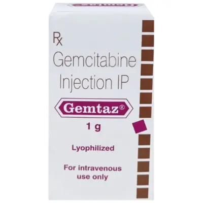
Gemcitabine + Nab-Paclitaxel:
-
Mechanism: Combines the antimetabolite Gemcitabine with Nab-Paclitaxel (paclitaxel bound to human albumin nanoparticles). The albumin carrier enhances drug transport across the vascular endothelium and helps break down the dense desmoplastic tumor stroma, allowing higher concentrations of gemcitabine to penetrate into cancer cells.
-
Clinical Role: An effective first-line regimen for patients who may not tolerate the gastrointestinal and neurological toxicities of mFOLFIRINOX.
-
Gemcitabine Monotherapy: Reserved as a gentle, palliative single-agent option for frail or elderly patients with compromised performance status.
2. Biomarker-Driven Targeted Therapies
Every patient diagnosed with advanced or metastatic pancreatic cancer should undergo Next-Generation Sequencing (NGS) and germline genetic testing:
-
PARP Inhibitors (Olaparib): For patients harboring germline BRCA1 or BRCA2 mutations whose cancer has not progressed after at least 16 weeks of first-line platinum-based chemotherapy. Olaparib acts via synthetic lethality, blocking alternative single-strand DNA repair pathways and inducing selective cell death, providing durable maintenance therapy.
-
Targeted KRAS G12D and G12C Inhibitors: While historically considered undruggable, small-molecule inhibitors targeting specific KRAS point mutations are advancing through clinical trials.
-
Immune Checkpoint Blockade: While standard mismatch repair-proficient (pMMR) pancreatic cancer does not respond to immunotherapy due to its non-immunogenic, cold tumor microenvironment, the rare subset (1% to 2%) of patients with MSI-High / dMMR pancreatic cancer achieve significant remissions with Pembrolizumab.
-
NTRK Gene Fusion Inhibitors: Patients harboring rare NTRK fusions can be treated with targeted oral inhibitors like Larotrectinib or Entrectinib.
Supportive Care, Symptom Control, and Quality of Life
Given the aggressive nature of pancreatic neoplasms, early integration of dedicated palliative and supportive care alongside active oncology therapies is essential:
-
Pancreatic Enzyme Replacement Therapy (PERT):
-
Management: Oral capsules containing standardized amounts of porcine lipase, protease, and amylase (e.g., Creon, Zenpep).
-
Administration: Capsules must be taken with the first bite of every meal and snack. Taking enzymes on an empty stomach or after finishing a meal renders them ineffective. Doses are adjusted based on meal fat content to eliminate steatorrhea, restore nutrient absorption, and stabilize body weight.
-
Biliary Decompression:
-
Endoscopic Stenting: For patients with obstructive jaundice from bile duct compression, a gastroenterologist performs an Endoscopic Retrograde Cholangiopancreatography (ERCP) to place an expandable Self-Expanding Metal Stent (SEMS) across the stricture into the duodenum, restoring natural bile flow, resolving jaundice, and clearing bilirubin so chemotherapy can proceed safely.
-
Celiac Plexus Neurolysis (CPN):
-
Severe Pain Control: For severe back pain caused by tumor invasion of retroperitoneal nerves, an interventional endoscopist can perform an ultrasound-guided injection of absolute alcohol or local anesthetics directly into the celiac nerve plexus. This disrupts visceral pain transmission, reducing dependence on high-dose opioid medications and decreasing opioid-related constipation and sedation.
-
Duodenal Stenting for Gastric Outlet Obstruction: If a tumor in the pancreatic head compresses the duodenum and prevents food from leaving the stomach, an endoscopic metal mesh stent or a surgical gastrojejunostomy bypass can restore oral intake.
High-Risk Screening and Prevention Strategies
While universal screening of the general population is not clinically recommended because the baseline population risk is low, structured surveillance programs are advised for defined, high-risk individuals:
1. High-Risk Surveillance Candidacy
Annual surveillance is recommended for individuals with:
-
Two or more first-degree relatives with pancreatic cancer, with at least one affected first-degree relative.
-
Three or more total relatives with pancreatic cancer.
-
Known pathogenic BRCA2, PALB2, or ATM mutations with a first- or second-degree relative with pancreatic cancer.
-
All individuals with Peutz-Jeghers Syndrome or FAMMM Syndrome (CDKN2A mutation), regardless of family history.
-
All individuals with documented Hereditary Pancreatitis (PRSS1 mutation).
2. Surveillance Protocol
-
Modalities: High-resolution Contrast-Enhanced MRI with MRCP alternating annually with Endoscopic Ultrasound (EUS), starting at age 50 (or 10 years earlier than the youngest family member diagnosed with pancreatic cancer), and starting at age 35 to 40 for Peutz-Jeghers and hereditary pancreatitis patients.
-
Surveillance Goal: Detecting high-grade precursor lesions (PanIN-3) or early, asymptomatic resectable Stage I adenocarcinomas before vascular involvement occurs.
Frequently Asked Questions
Can pancreatic cancer be cured completely?
Yes, pancreatic cancer can be cured if it is detected at an early stage when it is confined to the pancreas and can be removed with clear surgical margins. When a localized tumor is treated with complete surgical removal followed by a full course of modern adjuvant chemotherapy (such as mFOLFIRINOX), five-year survival rates range from 20% to 40%, with a subset of patients achieving long-term, permanent cure. However, because early pancreatic cancer often causes few symptoms, only roughly 15% to 20% of patients are candidates for surgery at initial diagnosis.
What is the most common early sign of pancreatic cancer?
In tumors located in the head of the pancreas, the most common early sign is painless obstructive jaundice—a yellowing of the skin and eyes accompanied by dark urine, pale or clay-colored stools, and itchy skin. For tumors located in the body or tail of the pancreas, the earliest signs are often a dull, deep ache in the upper abdomen that radiates into the mid-back, accompanied by unexplained weight loss and a loss of appetite.
Why is pancreatic cancer considered so difficult to treat?
Pancreatic cancer is difficult to treat due to three primary biological factors:
-
Late Detection: The pancreas sits deep in the abdomen, so tumors can grow without causing symptoms until they press on surrounding nerves, invade major blood vessels, or metastasize.
-
Dense Fibrous Tissue (Stroma): Pancreatic tumors are surrounded by a dense network of connective tissue that compresses local blood vessels, making it difficult for intravenous chemotherapy drugs to penetrate into the cancer cells.
-
Early Microscopic Spread: Even small pancreatic tumors tend to shed cells into regional lymph nodes and the bloodstream early, meaning systemic disease is often present even when scans show an apparently localized mass.
What is a Whipple procedure, and what organs are removed?
A Whipple procedure (pancreaticoduodenectomy) is a major surgical operation used to treat tumors in the head or uncinate process of the pancreas. During the surgery, a surgeon removes the head of the pancreas, the duodenum (the first section of the small intestine), the lower part of the common bile duct, the gallbladder, and nearby lymph nodes. The surgeon then reconnects the remaining part of the pancreas, the bile duct, and the stomach directly to the small intestine so food, bile, and digestive juices can continue to flow.
Can diabetes be an early warning sign of pancreatic cancer?
Yes. The sudden onset of type 2 diabetes in an adult over age 50 who does not have typical risk factors (such as obesity or a strong family history of diabetes), or a sudden, unexplained loss of blood sugar control in someone whose diabetes was previously stable, can be an early warning sign. Tumors can disrupt normal insulin secretion or produce metabolic signaling molecules that cause tissue insulin resistance months before gastrointestinal symptoms appear.
What should I do if my back pain gets worse when I lie flat?
Back pain that sits in the upper or middle back, feels like a deep ache, worsens when lying flat on your back, and eases slightly when curling forward or sitting up is a characteristic sign of retroperitoneal irritation. While frequently caused by spinal, muscular, or digestive issues, it should be evaluated by a physician—especially if accompanied by unexpected weight loss, nausea, or changes in stool color. A contrast-enhanced abdominal CT scan can evaluate the pancreas and retroperitoneum.
What are pancreatic enzyme capsules (PERT), and why are they needed?
When a tumor blocks the pancreatic duct, or after parts of the pancreas are surgically removed, the body cannot produce or release enough digestive enzymes to break down food. This leads to exocrine pancreatic insufficiency (EPI), causing greasy, loose, floating stools, severe gas, and progressive weight loss. Pancreatic Enzyme Replacement Therapy (such as Creon or Zenpep) provides prescription capsules containing natural enzymes that must be swallowed with food to break down nutrients and restore healthy digestion.
Is genetic testing recommended for pancreatic cancer patients?
Yes. Clinical guidelines recommend that all individuals diagnosed with pancreatic cancer undergo both germline genetic testing (testing inherited DNA from a blood or saliva sample) and somatic tumor profiling (next-generation sequencing on biopsy tissue). Identifying specific genetic variants—such as BRCA1, BRCA2, PALB2, or Mismatch Repair gene defects—can open up targeted therapy options (like PARP inhibitors or immunotherapy) and provides valuable information for family members who may benefit from early surveillance programs.
Medical Authority and Clinical Reference Links
-
National Comprehensive Cancer Network (NCCN):
NCCN Clinical Practice Guidelines in Oncology: Pancreatic Adenocarcinoma.
Consensus clinical practice guidelines for high-resolution CT imaging, resectability criteria, vascular resection standards, and systemic chemotherapy:
-
American Society of Clinical Oncology (ASCO):
ASCO Clinical Practice Guidelines: Locally Advanced and Metastatic Pancreatic Cancer.
Evidence-based consensus recommendations covering frontline chemotherapy combinations, germline testing, and supportive palliative care:
-
European Society for Medical Oncology (ESMO):
ESMO Clinical Practice Guidelines for Diagnosis, Treatment, and Follow-up of Pancreatic Cancer.
International recommendations covering diagnostic pathways, surgical margin evaluation, and targeted therapies:
-
International Association of Pancreatology (IAP) & American Pancreatic Association (APA):
Evidence-Based Guidelines for the Management of Intraductal Papillary Mucinous Neoplasms (IPMN) and Pancreatic Cystic Lesions:
-
Pancreatic Cancer Action Network (PanCAN):
Clinical Compendia: Precision Promise, Biomarker Testing Directory, and Patient Care Protocols: